


Rhinology
Vol. 46: Issue 4 - August 2026
Radiologic and cadaveric spatial relation between nasopalatine canal and the middle turbinate axilla
Authors
Octavio Garaycochea
, Isam Alobid
, Carla Rodríguez-Zanetti
 , Laura Pujol-Vilà
, Marta Calvo-Imirazaldu
, Secundino Fernandez
, Laura Pujol-Vilà
, Marta Calvo-Imirazaldu
, Secundino Fernandez
Summary
Cover Image
Objective. This study aims to describe the spatial relation (SR) between the nasopalatine canal (NPC) and the septal projection of the middle turbinate axilla (MTA-SP), 2 anatomical landmarks used in rhinology.
Methods. A retrospective study was conducted in patients who underwent a cone bean computed tomography. Two radiological measurements were assessed by 2 examiners: the distance between the perpendicular line corresponding to the MTA-SP to the posterior border of the NPC and the distance between the anterior and posterior border of the NPC. Also, an anatomical dissection was performed on a cadaveric head and NPC was cannulated to visually assess the SR through a radiological reconstruction.
Results. Among 30 patients, 14 females and 16 males with a mean age of 46.6 (SD 18.8) years, the mean distance between the perpendicular line corresponding to the MTA-SP to the posterior border of the NPC was -0.08 mm (SD 2.4 mm) and the mean distance between the anterior and posterior border of the NSP was 2.7 mm (SD 1.4 mm).
Conclusions. The septal projection of the middle turbinate axilla is above and slightly in front to the NPC, at a mean distance of 0.08 mm. Understanding this relationship provides essential information about structure and spatial orientation of the nasal cavity.
Introduction
The nasal septum (NS) is an osseocartilaginous structure located in the midline of the nasal fossa (NF) forming the medial wall of the nasal cavities. It has a primary role in respiratory physiology and nasal aesthetics 1. At the lower part of the NS, a bony passage provides a direct communication between the oral and nasal cavity (NC) opening into the nasal floor (Stenson foramina) at approximately 20 mm from the columella and 15 mm from the anterior nasal spine 2,3. This passage, known as the nasopalatine canal (NPC), also anterior palatine or incisive canal, contains the terminal branch of the greater palatine artery (also nasopalatine artery), the nasopalatine nerve, and the nasopalatine duct along with small salivary glands, adipose and connective tissue 4. The oral opening of the NPC has traditionally been considered more clinically relevant, particularly in orthodontic treatment, compared to its nasal opening 5. However, in recent years, the nasal opening, observable in 94% of cases and exhibiting various morphological presentations (round, oval, spindle or tubular), has garnered increasing attention from a rhinological perspective 6. It has been identified as a key anatomical landmark for topical vasoconstriction and local infiltration 7, for delimiting the anterior portion of the NS 8, and for evaluating the use of greater palatine artery flap as a surgical option for closure of nasal septum perforations 3.
Parallel to the NS in each NF is the turbinate wall, formed by the inferior, middle (MT) and superior turbinates. This structure is part of the lateral nasal wall and separates the nasal fossa from the adjacent paranasal sinuses 9. The MT can be divided into 3 segments. The first, anterior sagittal-oriented segment (lamina recurvata), is parallel to the lamina papyracea laterally and the NS medially, perpendicular to the skull base and superiorly attached to it. The second, coronal-oriented segment, corresponds to the sigma and concave/convex plate of the basal lamella. The third, posterior transversely-oriented, corresponds to the horizontal plate of the basal lamella, is parallel to the nasal floor and skull base, and attached laterally to the crista ethmoidalis of the perpendicular process of the palatine bone 10. The anterior end of the first segment is attached laterally to the nasal surface of the maxillary bone’s frontal process, forming the so-called “middle turbinate axilla” (MTA). This attachment point, and its septal projection (MTA-SP) have been widely used as a stable anatomic landmark in endoscopic surgery 11-13.
Understanding how objects or points are positioned in space relative to another is defined as spatial relation (SR). It involves the ability to comprehend and mentally manipulate positions, directions and distances between points within a defined space. The NPC and the MTA are 2 bony anatomical landmarks – one inferiorly at the septal level and the other superiorly on lateral wall – that can be assessed clinically and radiologically. Since the SR between different anatomical landmarks can improve the anatomical orientation and help define and segment the structure of the NC, the aim of this study was to determine the SR between the NPC and the MTA-SP.
Materials and methods
Radiologic examination
A retrospective study was conducted at the Otorhinolaryngology and Head and Neck Surgery and Radiology Departments of the University Clinic of Navarre with the approval from the Institutional Ethical Committee. The study included cone beam computed tomography (CBCT) images of the NC and paranasal sinuses of patients assessed in the Rhinology Unit from May 2024 to July 2024. CBCT assessment was not routinely made and only performed when indicated based on each patient’s situation. Only patients without signs of pathology at the level of the nasal floor, inferior meatus, middle meatus and ethmoidal region were included. Patients with history of nasal trauma, dacryocystorhinostomy, septal/sinus/maxillofacial surgery or undergoing orthodontic treatment were excluded.
Using multiplanar reconstructions, the projection of the MTA-SP was first identified in the axial plane as previously described by Gras-Cabrerizo et al. 14. Once this point was localised and centred at the level of the NPC using the coronal plane as a reference, a perpendicular line was drawn in the sagittal plane form the MTA-SP to the nasal floor, in the slice where the anterior and posterior borders of the NPC were most clearly identifiable. Two radiological measurements were obtained from the sagittal slice using the ruler tool of the software: 1) distance between the perpendicular line corresponding to the MTA-SP to the posterior border of the NPC (when anterior to it the value was negative, while if posterior the assigned value was positive); 2) distance between the anterior and posterior border of the NPC in its nasal opening (Fig. 1).
Radiological measurements for each CBCT scan were independently performed by 2 examiners and intraclass correlation coefficient (ICC) was calculated to assess the consistency between their results. All measurements were included in the analysis. A Shapiro-Wilk test indicated that data distribution for both measures 1 and 2 was not normally distributed. A Mann-Whitney U test was conducted to evaluate differences between sex and measurements, and Spearman’s correlation was used to assess the relationship between age and measurements.
Anatomic examination
A fresh-frozen cadaveric head of an adult male specimen from the University of Navarre School of Medicine’s Anatomical Donor Program was used for the anatomic examination.
To evaluate the SR between the NPC and MTA-SP from an endoscopic perspective, the nasal opening of the NPC in the left NC was identified using a 0° lens. Once located, it was visualised from the NC using 30°, 70°, and 120° lenses. Subsequently, the oral mucosa at the level of the hard palate beneath the incisive papilla was dissected to identify the oral opening of the NPC. Once identified, the canal was accessed with a 90° angled probe until it was visualised within the NC.
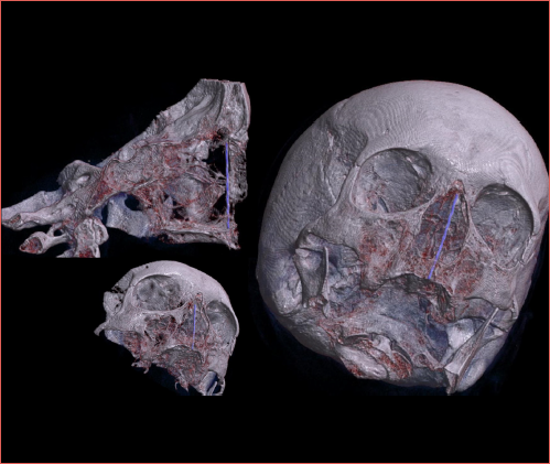
An open rhinoplasty was then performed through a transcolumellar approach to fully expose the nasal bones. Once exposed, an opening of approximately 10 mm was made with a cutting burr on the lateral margin of the left nasal bone, through which a 120° lens was introduced to obtain a complete view of the NC and the nasal opening of the NPC from the MTA. Finally, the NPC was cannulated with a rigid metal stent until reaching the approximate location of the MTA-SP. After the anatomical dissection was completed, a non-contrast CT of the specimen was performed using a single-source third-generation CT scanner (SOMATOM X, Siemens Healthcare) (Cover figure and Figure 2).
Results
Thirty CBCT facial scans were analysed, 14 females and 16 males (mean age, 46.6 years; SD, 18.8). ICC between both measures indicated a good reliability for both measurements (0.834 and 0.873, respectively). The mean distance between the perpendicular line corresponding to de MTA-SP to the posterior border of the NPC was -0.08 mm (SD 2.4 mm). In 55% of cases, the line corresponding to the MTA-SP coincided exactly with the NPC, in 15% with the anterior border, in 25% within the NPC and in 15% with the posterior border (Fig. 3). There was no significant difference between sex and measure 1 (p = 0.49). There was a significant difference between age and measurement 1 (p = 0.01) with a weak correlation (R = 0.17).
The mean distance between the anterior and posterior border of the NPC was 2.7 mm (SD 1.4 mm). There was a significant difference between sex and measurement 2 (p = 0.04), with a mean 18.5 mm in males and 11.9 mm in females. There was no significant difference between age and measurement 2 (p = 0.13). Images of the anatomic examination are described in Figure 2 and Cover figure.
Discussion
The SR between anatomical structures has been previously studied and demonstrated to be of significant clinical and surgical relevance 15. The main finding of this study is the close SR between the MTA-SP and NPC: the MTA-SP is above and slightly in front to the NPC, at a mean distance of 0.08 mm (SD 2.4 mm). Understanding this relationship provides essential information to rhinologists about the structure and spatial orientation within the NC. This is particularly important, as during nasal endoscopic examination or surgery with a 0° endoscope, the angle of view forms a 20-to-25° tilt relative to the horizontal plane, making the MTA appear more posterior than the NPC.
In fact, the MTA-SP is located superior and slightly anterior to the NPC. Knowing this alignment helps the surgeon estimate the position of instruments or the endoscope within the NC, especially in narrow spaces like the nasal floor or the anterior skull base. For example, if the endoscope moves higher within the fossa and the MTA becomes visible, the surgeon can infer the superior positioning relative to the NPC. These anatomical landmarks and their SR can help mitigate challenges of estimating sizes, distances, and the limited depth of field in nasal endoscopy 16.
This SR can be explained from an embryological perspective. The NPC is located at the junction of the primary (PP) and secondary palate (SP). Although recent studies suggest that this canal does not originate at the fusion area but adjacent to it (within the PP), it structurally separates both palates 17. During the embryological development of the NC, the olfactory placodes divide into medial and lateral olfactory processes. According to the evo-devo theory, the medial nasal processes fuse at the midline to form the intermaxillary process, which subsequently gives rise to the SLC, while the lateral olfactory processes form the lobular cartilage. The PP initially serves as the floor of the deepening olfactory until it fuses with the SP (in the classical theory, the intermaxillary process gives rise to the primary palate) 18.
Above the SLC, the nasal bones fuse at the midline, projecting caudally under the frontal bone’s glabella. The maxillary bone fuses with the lateral border of the nasal bone at its anterior cranial edge and with the nasal process of the frontal bone at its anterior superior edge. The union point between the frontal process of the maxillary bone and the nasal process of the frontal bone, adjacent to the orbit, corresponds to the region in the NC where the MTA is located (the inferomedial aspect of the floor of the frontal sinus). Caudally, the maxillary bones meet at the midline under the frontonasal process to form the SP, which fuses anteriorly with the PP, where the NPC will be formed. From an embryologic standpoint, both the NPC and the MTA are situated at the anterior portion of the maxillary swellings, one above the other, which relates to our findings.
This study also addresses the topic of anatomic nomenclature. Currently, there is a lack of consensus and limited applicability of Cottle’s classification in clinical practice 19, mainly due to inconsistencies in the literature regarding area definitions and artificial or non-anatomically defined boundaries 9. A new classification of NS areas has been proposed by our group, built upon Cottle’s concept of dividing the NS into specific regions and incorporating nasal functional areas described by Mlynski and Huizing 8. This classification divides the NS into 3 distinct areas, using 2 anatomical landmarks that can be clinically and radiologically assessed: NPC and posterior edge of the hard palate. It is based on the embryology, vascular supply, and surgical anatomy of the NS. An anatomical reference for the NPC in the upper region of the nasal fossa – both medially (MTA-SP) and laterally (MTA) – can help in the spatial localisation of the anterior septal area described in this classification.
Moreover, the SR between the NPC and the MTA suggests that the classification proposed by our group may complement, to some extent, the division of the lateral nasal wall proposed by Bodino-Jankoski et al. 9, which divides into pre-turbinal and turbinal wall by an imaginary line perpendicular to the NC floor. This line extends superiorly at the level of the MTA and inferiorly just anterior to the head of the inferior turbinate. Anterior to this line, the pre-turbinal lateral wall includes the lateral wall of the nostril, the vestibule, the nasal valve area, and Cottle’s attic area, which correspond to the external nose and face. These structures are also encompassed within the projection of Area I of our septal classification on the lateral nasal wall. The only difference is that, at the anterior-inferior level, the head of the inferior turbinate is also included in the projection of Area I; however, according to Bodino-Jankoski, it is considered part of the turbinal wall. Although both classification systems segment parallel – the NS and the lateral wall – that do not necessarily align due to their differences in function, size, and structure, their projection notably converge at a common anatomical landmark: the MTA and its septal projection. Regarding where the head of the inferior turbinate is located, we consider it more appropriate to classify it as part of the anterior nasal region.
This SR is not only valuable from an anatomical perspective, it can play a key role in surgical navigation. The MTA serves as an important landmark in the lateral wall, guiding various surgical approaches. In frontal sinus surgery, it facilitates access to the frontal sinus from the lateral wall, and the mucosa above it is involved in mucosal flaps in Draf IIb or Draf III procedures 20. Laterally, it helps locate the lacrimal sac during endonasal dacryocystorhinostomy and serves as a boundary for transnasal endoscopic partial maxillectomies, where type 3B may partially or completely remove the ascending-frontal branch of the maxillary bone) 12,21. Additionally, the MTA provides a reference point for accessing the frontal recess 22, and assists in the localisation of the anterior ethmoid artery (AEA), which is positioned at an average distance of 21 mm 23. Although different patterns of pneumatisation of the agger nasi cells can occur above the anterior end of the MT, this cell is closely related to the MTA 24. Furthermore, the MTA-SP is approximately at 7.3 mm anteriorly (SD 0.9) to the entry point of the nasal branch of the AEA into the NS, which is useful to know when performing AEA flaps 13. The MTA-SP is also linked to Stamm’s S-point, which corresponds to the “axilla” projection of the MT, located posterior to the septal body 25.
Regarding the NPC size, our results align with the existing literature. Although the size of the NPC depends from its shape and angulation, a diameter averaging less than 6 mm is considered normal. Difference between sexes has also been previously described, with larger diameters in men. In our results, no relationship was found between age and canal size, although it has been reported that canal width increases with age.
The primary limitation of this study is that the anatomical analysis was conducted on a single specimen, and the radiological assessment included only 30 subjects, limiting the generalisability of the findings to broader population. Furthermore, other anatomical factors of the NPC that influence its size and, therefore, the location of the posterior edge, such as its shape and angulation, were not considered.
Conclusions
The main conclusion of this study is that the middle turbinate axilla and its septal projection is above and slightly in front to the nasopalatine canal, at a mean distance of 0.08 mm. Understanding this relationship provides essential information to rhinologists about structures and spatial orientation within the NC during endoscopic surgery.
Conflict of interest statement
The authors declare no conflict of interest.
Funding
This research did not receive any specific grant from funding agencies in the public, commercial, or not-for-profit sectors.
Author contributions
SF, OG, IA, CRZ, LP: conceptualization; SF, OG, IA, CR-Z, MC, LP: methodology and materials, investigation; MC: software; SF, OG: formal analysis; CR-Z, OG: data curation; OG: writing – original draft preparation, project administration; CR-Z, LP: writing – review and editing; IA, SF: supervision. All authors have read and agreed to the published version of the manuscript.
Ethical consideration
This study as approved by CEI: investigation ethics committee of the Clinica Universidad de Navarra (protocol number: 2024.203, 19/08/2024). The research was conducted ethically, with all study procedures being performed in accordance with the requirements of the World Medical Association’s Declaration of Helsinki.
History
Received: April 14, 2025
Accepted: February 20, 2026
Figures and tables
Figure 1. Examples of radiological measurements. Red line – Measurement 1: distance between the perpendicular line corresponding to the MTA-SP to the posterior border of the NPC. Green line – Measurement 2: distance between the anterior and posterior border of the NSP. MTA-SP: septal projection of the middle turbinate axilla; PB: posterior border of the nasopalatine canal.
Figure 2. Anatomic examination. A) Oral opening of the NPC; B) Nasal opening of the NPC; C) Optic lens introduction through the nasal bone; D) Cannulation of the NPC, nasal floor perspective; E) Cannulation of the NPC, superior perspective; F) Coronal view of the specimen with the canula; G) Sagittal view of the specimen with the canula and radiological measurements. Black arrow: opening of the NPC; white arrow: optic lens; black arrowhead: canula; white asterisk: nasal vestibule; black asterisk: middle turbinate; NS: nasal septum; IT: inferior turbinate.
Figure 3. Spatial relation between the NPC and the septal projection of the middle turbinate axilla. The posterior border of the NPC is located at point 0.
References
- Huizing E, de Groot J. Functional Reconstructive Nasal Surgery. Thieme; 2003. doi:https://doi.org/10.1055/b-002-40814
- Lake S, Iwanaga J, Kikuta S. The incisive canal: a comprehensive review. Cureus. 2018;10. doi:https://doi.org/10.7759/cureus.3069
- Santamaría-Gadea A, Vaca M, de los Santos G. Greater palatine artery pedicled flap for nasal septal perforation repair: radiological study and case series. Eur Arch Otorhinolaryngol. 2021;278:2115-2121. doi:https://doi.org/10.1007/s00405-020-06230-1
- Fernández-Alonso A, Antonio Suárez-Quintanilla J, Muinelo-Lorenzo J. Critical anatomic region of nasopalatine canal based on tridimensional analysis: cone beam computed tomography. Sci Rep. 2015;5. doi:https://doi.org/10.1038/srep12568
- Tilen R, Patcas R, Bornstein M. The nasopalatine canal, a limiting factor for temporary anchorage devices: a cone beam computed tomography data study. Eur J Orthod. 2017;39:646-653. doi:https://doi.org/10.1093/ejo/cjx022
- Jacob S, Zelano B, Gungor A. Location and gross morphology of the nasopalatine duct in human adults. Arch Otolaryngol Head Neck Surg. 2000;126:741-748. doi:https://doi.org/10.1001/archotol.126.6.741
- Williams W, Ghorayeb B. Incisive canal and pterygopalatine fossa injection for hemostasis in septorhinoplasty. Laryngoscope. 1990;100:1245-1247. doi:https://doi.org/10.1288/00005537-199011000-00022
- Garaycochea O, Alobid I. On the Cottle areas and the proposal for a new classification of septal areas. Rhinology. 2023;61:190-192. doi:https://doi.org/10.4193/Rhin22.229
- Bodino C. Surgical anatomy of the turbinal wall of the ethmoidal labyrinth. Rhinology. 2004;42:73-80. doi:https://doi.org/10.4193/Rhin
- Kamel R. Understanding the complex anatomy of the middle turbinate via educational origami. Rhinol Online. 2018;1:154-159. doi:https://doi.org/10.4193/RHINOL/18.072
- Kosugi E, Balsalobre L, Mangussi-Gomes J. Breaking paradigms in severe epistaxis: the importance of looking for the S-point. Braz J Otorhinolaryngol. 2018;84:290-297. doi:https://doi.org/10.1016/j.bjorl.2017.12.007
- Shams P, Wormald P, Selva D. Anatomical landmarks of the lateral nasal wall: implications for endonasal lacrimal surgery. Curr Opin Ophthalmol. 2015;26:408-415. doi:https://doi.org/10.1097/ICU.0000000000000160
- Naidu L, Sibiya L, Aladeyelu O. Anatomical landmarks for localisation of the anterior ethmoidal artery: a combined radiological and cadaveric (endoscopic) study. Surg Radiol Anat. 2023;45:545-554. doi:https://doi.org/10.1007/s00276-023-03122-x
- Gras-Cabrerizo J, García-Garrigós E, Ademá-Alcover J. A unilateral septal flap based on the anterior ethmoidal artery (Castelnuovo’s flap): CT cadaver study. Surg Radiol Anat. 2016;38:723-728. doi:https://doi.org/10.1007/s00276-015-1610-9
- Tham A, Purchase R, Kelly J. The relation of the coracoid process to the glenoid: an anatomic study. Arthroscopy. 2009;25:846-848. doi:https://doi.org/10.1016/j.arthro.2009.01.022
- Weber R, Hosemann W. Comprehensive review on endonasal endoscopic sinus surgery. GMS Curr Top Otorhinolaryngol Head Neck Surg. 2015;14. doi:https://doi.org/10.3205/cto000123
- Radlanski R, Emmerich S, Renz H. Prenatal morphogenesis of the human incisive canal. Anat Embryol (Berl). 2004;208:265-271. doi:https://doi.org/10.1007/s00429-004-0389-y
- Jankowski R, ed. The Evo-Devo Origin of the Nose, Anterior Skull Base and Midface.; 2013. doi:https://doi.org/10.1007/978-2-8178-0422-4_8
- Huizing E. Incorrect terminology in nasal anatomy and surgery, suggestions for improvement. Rhinology. 2003;41:129-133.
- Fiorini F, Nogueira C, Verillaud B. Value of septoturbinal flap in the frontal sinus drill-out type IIb according to Draf. Laryngoscope. 2016;126:2428-2432. doi:https://doi.org/10.1002/lary.25947
- Turri-Zanoni M, Battaglia P, Karligkiotis A. Transnasal endoscopic partial maxillectomy: operative nuances and proposal for a comprehensive classification system based on 1378 cases. Head Neck. 2017;39:754-766. doi:https://doi.org/10.1002/hed.24676
- Wormald P. The axillary flap approach to the frontal recess. Laryngoscope. 2012;112:494-499. doi:https://doi.org/10.1097/00005537-200203000-00016
- Lee W, Ku P, Van Hasselt C. New guidelines for endoscopic localization of the anterior ethmoidal artery: a cadaveric study. Laryngoscope. 2000;110:1173-1178. doi:https://doi.org/10.1097/00005537-200007000-00020
- Rusu M, Sava C, Ilie A. Agger nasi cells versus lacrimal cells and uncinate bullae in cone-beam computed tomography. Ear Nose Throat J. 2019;98:334-339. doi:https://doi.org/10.1177/0145561319840836
- Kosugi E, Balsalobre L, Stamm A. Refractory epistaxis and Stamm’s S-point. Curr Opin Otolaryngol Head Neck Surg. 2022;30:13-18. doi:https://doi.org/10.1097/MOO.0000000000000773
Downloads
License

This work is licensed under a Creative Commons Attribution-NonCommercial-NoDerivatives 4.0 International License.
Copyright
Copyright (c) 2026 Società Italiana di Otorinolaringoiatria e chirurgia cervico facciale
How to Cite
- Abstract viewed - 267 times
- PDF downloaded - 80 times