Otology
Vol. 45: Issue 3 (Suppl. 1) - June 2025
Stapes revision surgery: intraoperative findings and audiological results. A multicentric study
Summary
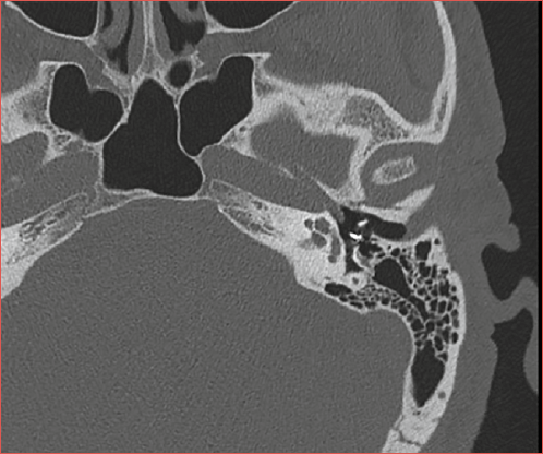
Cover Image
Objective. To explore the results of revision stapes surgery within a multicentric cohort, focusing on hearing improvements and correlation with the type of postoperative hearing loss experienced and related findings during the revision procedure.
Methods. A retrospective study of 308 consecutive revision stapes surgeries performed ay 5 Otorhinolaryngologic Units in Pisa, Turin, Pavia, Piacenza, and Rome between 2010 and 2023 was accomplished.
Results. The most frequent causes leading to revision stapes surgery were prosthesis dislocation (56.1%), use of a short prosthesis during primary surgery (13.9%), and an eroded incus (17.2%). The median air conduction threshold significantly improved after revision surgery, while the bone conduction threshold remained stable.
Conclusions. Revision stapes surgery effectively improves hearing in patients with unsuccessful initial operations, with a significant reduction in the air-bone gap (ABG). The audiological results in our patients are favourable. ABG closure within 10 dB was achieved in 57.2% of cases and within 20 dB in 76% of cases. Stapes revision surgery is feasible and provides an acceptable success rate.
Introduction
Since the mid-20th century, surgical intervention has been the standard treatment for otosclerosis-related hearing impairment 1. Stapes surgery is effective in reducing the air-bone gap (ABG) to less than 10 dB in nearly 90% of cases 2. However, some authors have pointed out that up to 20% of patients may require a second surgery due to persistent or recurrent hearing loss or vertigo 3-7. Immediate failures leading to continued hearing loss can arise from issues such as improper placement of the prosthesis or other causes like superior semicircular canal dehiscence or ossicular chain problems 8. On the other hand, delayed failures, presenting as either gradual or sudden recurrence of hearing loss, can be due to reasons such as prosthesis dislocation, erosion of the incus, regrowth of the footplate, or scarring 8.
Revision operations for otosclerosis generally yield less favourable results than primary surgery, with successful ABG reduction observed in 40% to 78% of cases during the first surgical revision and in 21% during the second revision 9-12. Revision surgery is also reported to be more frequently accompanied by complications like sensorineural hearing loss and vertigo 9-12. Moreover, the risk of deafness appears to be 5 times higher after revision surgery compared to primary surgery (0.5% or below) 12. Nevertheless, some recent studies have begun to question this trend, pointing out the variability in outcomes and the scarcity of research into the causes for revision surgery and their impact on success rates 13,14.
Surgery remains the preferred treatment for otosclerosis, including revision cases. Hearing aids, despite their utility, offer a lower quality of life compared to surgery and come at a higher cost 15. Bone-implanted prostheses serve as a third option following surgical interventions, including stapes revision surgery and the use of conventional prostheses 16.
The aim of this study was to explore the results of revision stapes surgery within a multicentric cohort. We focused on hearing improvements and their correlation with the type of postoperative hearing loss experienced and the related findings during the revision procedure. This investigation aims to provide better guidance for patients undergoing revision stapes surgery following an unsuccessful initial operation.
Materials and methods
Patients
All 308 consecutive revision stapes surgeries performed at 5 Otorhinolaryngologic Units in Pisa, Turin, Pavia, Piacenza, and Rome between 2010 and 2023 were enrolled in this retrospective study. These surgeries were carried out on 283 subjects, as 15 underwent 2 revision procedures, and 5 underwent 3 revisions.
Medical charts were analysed for demographic data and medical history. The surgical report of primary surgery was reviewed to define the operative technique employed (stapedotomy or stapedectomy). Intraoperative findings during the revision surgery (e.g., erosion of the long process of the incus, perilymphatic fistula, malleus fixation) were recorded, and the data were correlated with postoperative audiological results. For patients who underwent high-resolution computed tomography (HRCT) before the revision surgery, radiological findings were evaluated and compared with intraoperative findings. Pure tone audiometry was performed pre- and postoperatively in a soundproof cabin according to the ISO 8253-1 and 8253-3 standards.
Surgical procedure
In almost all cases, the revision surgery was performed using a transcanal approach under local anaesthesia, as in primary surgery 17. Only upon the patient’s request, due to anxiety or other reasons, was the procedure performed under general anaesthesia. During revision surgery, the malleus and incus were visually inspected, and their mobility assessed by gentle palpation. The connection of the prosthesis to the incus or malleus and its position in the niche of the oval window were checked. Scars or bridles were gently removed with a tip; laser was used by some surgeons within our group for this purpose. No stapedectomy was reported in the primary surgery; instead, all cases involved a stapedotomy followed by placement of a piston between the incus and the platinar hole. During revision surgery, the original stapedotomy fenestra was inspected. Anatomic/pathologic irregularities of the middle ear were found in only 72 cases (23.3%). In particular, facial nerve dehiscence was described in 13 (4.2%) patients, and obliteration of the oval window in 4 (1.3%).
During revision, the causes of hearing deterioration were identified and corrected. In case of prosthesis dislocation, a new footplate hole was made using a microdrill or laser, and a new prosthesis repositioned. If the stapedial piston prosthesis was too short, it was replaced by a prosthesis of adequate length. If the incus was eroded, the new stapedial prosthesis was attached to the residual incus or fixed by performing a malleostapedioplasty. In case of perilymphatic fistula, it was sealed using autologous fat on the oval window or perichondrium. A malleostapedioplasty with section of the head was performed in cases of malleus-incus ankylosis 18.
During primary surgery, 2 types of prostheses were predominantly used: platinum-Teflon in 62.3% and Teflon in 36.3% of cases. Other types of prostheses were rarely used, including nitinol (0.3%), titanium (0.3%), and titanium-Teflon (0.6%). Similarly, in revision surgery, platinum-Teflon prostheses were used in 58.4% and Teflon prostheses in 35% of cases, with other types being rarely employed: nitinol (0.6%), Teflon-titanium (4.8%), and titanium (0.6%).
Audiometric assessment
Audiometric evaluation included assessment of pre- and postoperative air-conduction (AC) thresholds (at 0.25, 0.5, 1, 2, 3, 4, and 8 kHz), bone-conduction (BC) thresholds (at 0.25, 0.5, 1, 2, 3, and 4 kHz), and ABG (at 0.5, 1, 2, and 3 kHz). Only AC and BC results obtained at 12 months after surgery were used for the calculation of the ABG and pure-tone averages (PTAs). Postoperative “dead ear” was defined as profound sensorineural hearing loss on all postoperative audiologic examinations. All patients had at least a 12-month audiological follow-up. Audiometry was reported according to the American Academy of Otolaryngology – Head and Neck Surgery guidelines 19.
Statistical analysis
A descriptive analysis of all variables, including quantitative and qualitative data, was reported. The normal distribution of numerical variables was assessed using the Shapiro-Wilk test and the Kolmogorov-Smirnov test. The numerical variables were not normally distributed. Subsequently, the variables were compared using suitable statistical methods, including the Mann-Whitney U test for non-parametric data and the Chi-square test for categorical variables. The correlation between variables was determined using Spearman’s rank correlation for univariate analysis and linear regression for multivariate analysis. The software used for the analysis was the SPSS version 23 (IBM Corp., Chicago, IL, USA).
Results
Patients
The cohort comprised 202 females (65.5%) and 106 males (34.5%), with a male-to-female ratio of 1:1.9. The mean age of the cohort was 52 years (± 11.5, range 23-80). Of all surgical procedures, 283 (91.9%) were primary revisions, 20 (6.5%) were secondary revisions, and 5 (1.6%) were tertiary revisions. The indications for revision surgeries were either hearing loss or vertigo. Revision surgery was recommended in 295 cases (95.8%) for persistent or recurrent hearing loss characterised by an average ABG of 4 frequencies above 20 dB, and in 9 cases (2.9%) for vertigo, imbalance, and/or signs of perilymphatic fistula. Moreover, the surgical procedure was interrupted in 4 cases (1.3%).
The surgical procedures were mostly performed under local anaesthesia (94.2% of cases) and less frequently under general anaesthesia (5.8%), according to the patient’s and surgeon’s preferences. Local anaesthesia was the preferred option for individuals with vertigo.
Before the revision surgery, CT was conducted in 63 subjects, constituting 20.4% of the entire study group. CT accurately identified dislocated prostheses in about half of these cases (41.2%) and a short prosthesis in 8 cases (12.6%). In the remaining instances (46.1%), scan failed to identify the underlying cause necessitating surgical revision. Therefore, the sensitivity of the test in our dataset is 53.9%, while the specificity cannot be calculated as the group lacked healthy subjects.
Intraoperative results
The causes of failure after primary surgery were intraoperatively evaluated. These findings are detailed in Table I. The most frequent causes leading to revision surgery were prosthesis dislocation (56.1%), eroded incus (17.2%), and use of a short prosthesis during primary surgery (13.9%). These 3 causes accounted for 87.2% of the reasons leading to revision in our population. In all cases, since the prosthesis was extruded from the footplate hole, it resulted in its re-ossification or closure by scar tissue.
Less frequent causes of primary surgery failure included hypomobility of the ossicular chain due to ankylosis or scar (8.4%), perilymphatic fistula (2.6%), prosthesis too long (0.3%), or interruption of the primary surgical procedure (1.3%).
The time of symptom onset after primary surgery was divided according to the surgical findings, as reported in Table I. Immediate surgical failures necessitating swift revision included a perilymphatic fistula (after 2 months) and a prosthesis that was too short (after 10 months). Conversely, other factors led to surgical revisions over a longer period, with incus erosion typically occurring after an extended period, averaging 114 months (data not shown).
The percentages of failure for the first operation, categorised by type of prosthesis used, are also presented in Table I. Prosthesis dislocation occurred in 47.4% of cases with platinum-Teflon prostheses and in 70.5% with Teflon prostheses. Incus erosion was observed in 21.3% of cases with platinum-Teflon prostheses and in approximately 10% with Teflon prostheses.
Audiometric results
The median AC and BC thresholds before stapedotomy were 55 dB and 27.8 dB, respectively. The median ABG was 27.2 dB. After stapedotomy, the median AC and BC thresholds were 52.5 dB and 29 dB, respectively. The median postoperative ABG was 23.4 dB. No statistically significant differences were found between pre- and post-stapedotomy AC and BC thresholds. However, the difference between pre- and post-stapedotomy ABG was statistically significant (p = 0.015).
Before revision surgery, the median AC and BC thresholds were 64.5 dB and 33.9 dB, respectively. The median pre-revision ABG was 30.4 dB. The median AC and BC thresholds after revision surgery were 46.3 dB and 32.8 dB, respectively. The median ABG was reduced to 13.3 dB. The post-revision AC thresholds were significantly better than the pre-revision ones (p < 0.001), without significant differences in pre- and post-revision BC thresholds. The difference between pre- and post-revision ABG was also significant (p < 0.001). These data are summarised in Table II.
The success of revision surgery can be expressed as the difference in ABG (ΔABG) before and after revision surgery, excluding patients with significant post-revision BC threshold decrement. The median value of this parameter was 16.2 dB. The post-revision ABG was better than 10 dB in 57.2% of cases and better than 20 dB in 76%, with only one case of cochlear damage, which was excluded from the statistical analysis.
Univariate analysis indicated a significant negative correlation between ΔABG and surgeries conducted under general anaesthesia (Spearman ρ = -0.177, p = 0.010). Furthermore, ΔABG was significantly negatively correlated with the number of days of hospitalisation (Spearman ρ = -0.372, p = 0.007). ΔABG was also positively correlated with higher pre-revision AC PTA (Spearman ρ = 0.234, p = 0.001) and negatively correlated with higher pre-revision BC PTA (Spearman ρ = -0.259, p < 0.001). ΔABG was also negatively correlated with post-revision AC PTA (Spearman ρ = -0.527, p < 0.001) and post-revision BC PTA (Spearman ρ = -0.184, p = 0.008).
Multivariate analysis using linear regression revealed that only the AC and BC thresholds retained a significant correlation with the difference in ABG before and after revision (p < 0.001). These data suggest that a greater pre-revision ABG may be correlated with a better ΔABG (Tab. III).
Discussion
The present study investigated the outcomes and challenges of secondary stapedotomy procedures in a retrospective series of 308 cases, revealing that significant improvements in hearing are possible with prompt revision surgery. In our series, dislocation of the prosthesis or its shortness accounted for approximately 70% of surgical revisions (Cover figure). Previous studies also indicate that issues related to the stapedial prosthesis are the most common reason for revision surgery, albeit with a lower prevalence (up to 60%) 10,12,20-22. In line with our data, displacement of the prosthesis was the most frequent cause 10,21. The high rate of prosthesis dislocation may be related to the type of prostheses used. Specifically, Teflon prostheses have been reported to have a dislocation rate of approximately 3% at 2 years after primary surgery 23. Also in our data set, Teflon prostheses showed a higher rate of dislocations compared to other prostheses (Tab. II). Although less frequent, the shortness of prosthesis used in primary surgery 21 remains a common cause of revision. These problems are more susceptible to repair with a higher success rate than other issues described below 10.
Erosion of the incus long process is another prevalent reason for undergoing revision surgery, with occurrences reported between 5% and 32% 9,10,21,22,24-26. In our series, this event occurred in 17.2% of cases. The literature extensively debates the possible causes of incus erosion. The long process of the incus receives blood supply from the incudal artery, which originates from the ossicular branch of the anterior tympanic artery and supplies the mucosal arteries and the nutrient foramen within the incus body. The lenticular process also receives blood through anastomoses from the arteries of the stapes tendon and posterior crural artery 27. It was previously thought that avascular necrosis could occur due to a compromised blood supply caused by an excessively tight crimping of the wire. However, Schuknecht refuted this, stating that the incus is well-supplied by a central nutrient vessel and a network of mucosal vessels sufficient to nourish the tip of the long process 27. More recent findings have indicated that the most part of nutrient foramina are in the upper two-thirds of the long process on its anteromedial side, a location generally unaffected by a crimped prosthesis 28. Nowadays, it is commonly accepted that incus necrosis is likely due to an overly loose crimp, allowing the prosthesis to continuously rub against and gradually wear down the long process 29. While this erosion is often associated with crimped wire, it can also occur with various other types of prostheses 25,30. The material of the prosthesis seems to favour a foreign body reaction. Platinum was suggested as a good option due to its malleability, but its use was associated with a higher occurrence of necrosis of the long process of the incus. Also in our data set, platinum-Teflon prostheses are associated with a higher rate of incus erosion than other prostheses. This increased rate of necrosis is believed to be associated with local toxicity or alterations in incus attachment 29,31. Data on rates of incus erosion and implant dislocation are not homogeneous in the literature (Tab. IV).
The variability in rates of incus erosion and prosthesis dislocation likely depends on the surgical technique used during the initial procedure and the type of prosthesis implemented. In our series, we observed a high percentage of prosthesis dislocations infrequently associated with incus erosion. The types of prostheses used in our series may explain the high rate of dislocation observed during revision surgery. Specifically, Teflon prostheses sometimes fail to adequately secure the long process of the incus, leading to dislocation without causing damage to the incus. Therefore, in our observations, a loosely crimped prosthesis that continuously moves does not gradually wear down the long process but instead dislodges from it. Our data do not align with Sakano’s claim that incus necrosis is due to a loose crimp that allows the prosthesis to continually rub and gradually erode the long process 29. Moreover, in our series, during the first surgical procedure, prostheses of different materials – Teflon, platinum-Teflon, titanium-Teflon, nitinol – were used, and prostheses in platinum-Teflon seemed to be associated with a higher prevalence of erosion of the incus.
Other causes of primary surgery failure, such as middle ear scarring, ankylosis, and perilymphatic fistula, were much less frequent. Adhesions and fibrous bands are occasionally observed. Some authors have reported that the incus and prosthesis may become immobilised due to dense fibrous tissue 6, but the occurrence of adhesions is more common in stapedectomy compared to stapedotomy (20.6% vs 7.9%). This finding supports our data, as stapedotomy is considered a less invasive technique 21. Schönfeld and collaborators emphasise the importance of avoiding damage to the mucosa around the oval window or promontory during initial surgery to prevent fibrosis that could adhere the prosthesis or incus to the promontory or tympanic membrane 34. In case of fibrous tissue and adherence, the use of a laser, particularly around the oval window, may facilitate a gentler removal of adhesions. One study highlighted that using an argon laser during revision surgeries increased successful outcomes from 70% to 91%, achieving an ABG within 10 dB. However, another more recent study found no significant impact on hearing improvement outcomes when comparing different energy levels of the laser or its use versus non-use in revision stapedectomy 34. In our series, scars around the piston and incus occurred in 6.4% of the surgical procedures, and we never used a laser to remove the fibrous bands.
To prevent persistent conductive hearing loss, it is critical to palpate the entire ossicular chain because a fixed malleus and/or incus may be congenital or caused by bone dust and fragments during primary surgery. This happened rarely (less than 4%) in our series, with 6 subjects affected (1.9% of cases) 6,12 and, unfortunately, there is no straightforward or uncomplicated way to resolve this problem. In 4 cases of our series, the surgeon performed a malleostapedotomy, cutting the head of the malleus with good but not optimal results.
Perilymphatic fistula is commonly encountered during revision surgeries performed due to the onset or persistence of vertigo. Typically, surgery is not indicated for sensorineural hearing loss following stapedectomy unless the patient experiences symptomatic vertigo suggestive of perilymphatic fistula. One study reported the incidence of delayed vertigo after stapedectomy as 0.5% 22. Another study indicated that perilymphatic fistula accounted for 9% of revision stapes surgeries, all associated with vertigo 22. This and other studies suggest that the presence of perilymphatic fistula is likely underestimated 21. Eight patients in our series had vertigo, and a perilymphatic fistula was found during surgical revision. The fistula was then sealed with perichondrium, leading to complete remission of symptoms. Literature data and our experience suggest that if a patient experiences vertigo (with or without sensorineural hearing loss), suspicion for perilymphatic fistula should be raised, and exploration should be performed.
The type of anaesthesia used for surgical revision is also important. Although any method of anaesthesia may be acceptable in primary surgery, local anaesthesia alone or local anaesthesia with sedation can have an advantage in revision surgery. If a patient suffers from dizziness while the surgeon manipulates or removes the previously placed prosthesis, this may indicate the presence of adhesions between the prosthesis and the sacculus. Without patient feedback, the surgeon may continue to manipulate or remove the prosthesis, placing the patient’s hearing at risk. Nevertheless, we found a significant positive correlation between ΔABG and general anaesthesia in univariate analysis (p = 0.010), although this was not confirmed in multivariate analysis.
The audiological results in our dataset are positive. ABG closure within 10 dB was achieved in 57.2% of cases and within 20 dB in 76%, with only one instance of profound sensorineural hearing loss. Revision surgery, however, remains challenging due to complications and its overall success rate, which is lower than that of primary surgery, even in the most experienced hands. Overall, ABG closure is 51-80% within 10 dB and 75-89.5% within 20 dB (Tab. V).
The risks of sensorineural hearing loss and vertigo are believed to increase with revision stapes surgery compared to primary stapes surgery. Historically, reported rates of sensorineural hearing loss after revision surgery have been as high as 20% 10. This increased risk may result from the distortion of normal anatomy due to prior surgery and unpredictable bony regrowth of the oval window. However, recent studies have shown a rate of sensorineural hearing loss of about 0-2.9% 11. Recent scientific papers indicate low rates of cochlear damage. This could be attributed to the fact that these studies discuss revisions of stapedotomies, which appear to have a lower incidence of cochlear damage compared to revisions of stapedectomies 37. Additionally, it is worth noting that very few authors have reported high rates of cochlear damage in the scientific literature 38. These rates fall within the reported ranges for stapes surgery, suggesting that routine revision stapes surgery is safe in experienced hands.
Univariate analysis of individual factors showed that the mean preoperative ABG is a positive prognostic factor for successful surgery, and this finding was confirmed by multivariate analysis. Sharaf et al. consider the ABG a predictive factor for success of revision stapedioplasty surgery 39. Additionally, previous stapedotomy was associated with higher success rates in revision surgery than previous partial or total stapedectomy 20.
Regarding intraoperative findings, prosthesis dislocation was associated with higher success rates than other primary causes of failure. Incus erosion and malleus-incus ankylosis were also associated with relatively high success rates 20. Moreover, the type of prosthesis placed during revision surgery was identified as a prognostic factor: a low rate of success was obtained for total ossicular prostheses compared with a high rate of success with other prostheses (pistons, wires, others) 40. Malleus-to-oval window interposition was associated with lower success rates than incus-to-oval window interposition 40.
Furthermore, the number of previous surgeries was identified as a negative prognostic factor 40. Considering all this data, the use of three variables – previous surgical technique, primary cause of failure, and type of prosthesis during revision surgery – can be used together in predicting success following stapes surgery 20. Differences in piston materials may confound comparisons of outcomes. Generally, it is acknowledged that there is no significant difference in hearing outcomes among the various types of pistons. Our study supports these findings. Approximately one-fourth of patients underwent revision stapedotomy using a laser. In our series, there were no significant differences between the laser and non-laser groups in terms of mean postoperative gap, the percentage of patients with a gap ≤ 10 dB, the mean change in sensorineural hearing loss, or the percentage of cases with improved hearing. Similar results have been described by other authors 40.
Surgical success can also be enhanced by understanding the cause necessitating revision surgery beforehand. Additionally, because some primary surgical procedures are performed at other institutions, information on primary surgical techniques and prostheses may be limited. In such cases, CT can be helpful. In our case series, CT identified the problem in more than half of cases. However, it is important to note that CT can miss imperceptible findings such as prosthesis displacement, incus erosion, or dehiscence of the superior semicircular canal. Therefore, the images should be reviewed collaboratively by radiologists and surgeons who are experienced in stapes surgery.
Conclusions
Revision stapedoplasty is feasible and yields good results with a low risk of cochlear damage. In our series, the primary intervention was always a stapedotomy. Therefore, future revisions will be predominantly revisions of stapedotomies. Intraoperatively, prosthesis dislocation was the most frequent finding, while incus erosion was infrequent. This can be attributed to the good performance of modern prostheses. The combination of primary stapedotomy and prosthesis dislocation leads to excellent surgical outcomes with an almost zero rate of cochlear damage.
Conflict of interest statement
The authors declare no conflict of interest.
Funding
This research did not receive any specific grant from funding agencies in the public, commercial, or not-for-profit sectors.
Author contributions
LB: conceptualization, writing – original draft preparation, writing – review and editing; LB, FL: methodology; FL: software; FF, LB, SB: validation; FL, LB: formal analysis; OM: investigation; all the authors: data curation, visualization; DC, AA, EC, AC: supervision; SB: project administration. All authors have read and agreed to the published version of the manuscript.
Ethical consideration
This study was approved by the Institutional Ethics Committee. Local Review Board do not release acceptance codes for retrospective studies based on clinical practice measures. The research was conducted ethically, with all study procedures being performed in accordance with the requirements of the World Medical Association’s Declaration of Helsinki.
Written informed consent was obtained from each participant/patient for study participation and data publication.
History
Received: January 17, 2025
Accepted: April 7, 2025
Figures and tables
| Main cause of failure | All surgical procedure (n = 308) | Prosthesis in platinum-Teflon (n = 192) | Prosthesis in Teflon (n = 112) |
|---|---|---|---|
| Dislocated prosthesis | 173 (56.1%) | 91 (47.4%) | 79 (70.5%) |
| Eroded incus | 53 (17.2%) | 41 (21.3%) | 11 (9.8%) |
| Short prosthesis | 43 (13.9%) | 24 (12.5%) | 19 (16.9%) |
| Long prosthesis | 1 (0.3%) | 1 (0.5%) | - |
| Malleus-incus ankylosis | 6 (1.9%) | 5 (2.6%) | 1 (0.8%) |
| Scar of the ossicular chain | 20 (6.4%) | 18 (9.3%) | 2 (1.7%) |
| Perilymphatic fistula | 8 (2.6%) | 8 (4.1%) | - |
| Surgery interrupted | 4 (1.3%) | 4 (2%) | - |
| Data are also grouped by the 2 more used types of prostheses in the study sample: platinum-Teflon and full Teflon; the data for the residual 4 prostheses (one in nitinol, one in titanium, and 2 in titanium-Teflon) are not reported. | |||
| Stapedoplasty | Preoperative (mean SD, range in dB) | Postoperative (mean SD, range in dB) | p value |
|---|---|---|---|
| Air conduction pure tone average (0.5-3 kHz) | 55.03 (± 14.57, 33.75-95) | 52.57(± 19.53, 15-100) | 0.396 |
| Bone conduction pure tone average (0.5-3 kHz) | 27.82 (± 12.41, 5-70) | 29.08 (± 12.86, 5-67.5) | 0.264 |
| ABG (0.5-3 kHz) | 27.21 (± 9.6, 6.25-50) | 23.47 (± 14.8, -3.75-56.25) | 0.015 |
| Revision surgery | |||
| Air conduction pure tone average (0.5-3 kHz) | 64.52 (± 17.02, 28.75-115) | 46.30 (± 20.59, 8.75-111.25) | 8.72 x 10⁻²⁴ |
| Bone conduction pure tone average (0.5-3 kHz) | 33.96 (± 14.1, 5-106.25) | 32.89 (± 15.16, 8.75-111.25) | 0.42 |
| ABG (0.5-3 kHZ) | 30.45 (± 12.06, -1.87-66.25) | 13.30 (± 12.42, 1.15-56.25) | 2.52 x 10-43 |
| Spearman’s rho | Multivariate linear regression analysis | ||||||
|---|---|---|---|---|---|---|---|
| Correlation coefficient | Sig. (2-tailed) | Standardised coefficient beta | t | Sig. | 95% CI - lower bound | 95% CI - upper bound | |
| Local anaesthesia “yes” | -0.177 | 0.010 | -0.030 | -1.085 | 0.279 | -3.913 | 1.131 |
| Use of laser “yes” | -0.085 | 0.127 | |||||
| Prosthesis width | -0.147 | 0.087 | |||||
| Prosthesis length | -0.025 | 0.775 | |||||
| Days of hospitalisation | -0.372 | 0.007 | 0.013 | 0.471 | 0.638 | -0.895 | 1.458 |
| Pre-revision air conduction PTA (0.5-3 kHz | 0.234 | 0.001 | 0.797 | 21.823 | 0.000 | 0.589 | 0.706 |
| Pre-revision bone conduction PTA (0.5-3 kHz) | -0.259 | 0.000 | -0.574 | -14.020 | 0.000 | -0.599 | -0.452 |
| Post-revision air conduction PTA (0.5-3 kHz) | -0.527 | 0.000 | -1.233 | -27.027 | 0.000 | -1.007 | -0.870 |
| Post-revision bone conduction PTA (0.5-3 kHz) | -0.184 | 0.008 | 0.795 | 16.155 | 0.000 | 0.720 | 0.919 |
| Difference in months between primary surgery and revision | 0.0878 | 0.328 | |||||
| References | Incus erosion | Displaced prosthesis | Short prosthesis |
|---|---|---|---|
| Bernardeschi et al. 32 | 42.1% | 20.6% | 3.9% |
| Blijleven et al. 9 | 5% | 27% | 15% |
| Fernandez et al. 14 | 32% | 26% | 21% |
| Kanona et al. 33 | 12% | 8% | 1% |
| Luryi et al. 8 | 38% | 43% | - |
| Schwam et al. 26 | 43.4% | 24.5% | - |
| Lundman et al. 12 | 35.6% | 48.2% | 3.1% |
| Vincent et al. 11 | 27.6% | 18.2% | 12.7% |
| Author | No. of cases | ABG ≤ 10 dB (%) | ABG ≤20 dB (%) | Dead ear (%) |
|---|---|---|---|---|
| Pedersen et al. 35 | 163 | 51 | 75 | 1.2 |
| Hammerschlag et al. 6 | 250 | 80 | 85 | 0 |
| De La Cruz et al. 40 | 356 | 60 | 78 | 1.4 |
| Lippy et al. 21 | 483 | 71 | 86 | 0.002 |
| Gros et al. 10 | 63 | 52.4 | 79.4 | 1.6 |
| Schmid et al. 36 | 172 | 55 | 84 | 1.2 |
| Vincent et al. 11 | 652 | 63 | 75 | 2.9 |
| Bernardeschi et al. 32 | 102 | 60 | 85 | 2 |
| Fernandez et al. 14 | 34 | 68.5 | 89.5 | 0 |
| Our data | 308 | 57.2 | 76 | 0.3 |
References
- House H. Footplate surgery in otosclerosis. J Laryngol Otol. 1962;76:73-86. doi:https://doi.org/10.1017/s0022215100059065
- Kishimoto M, Ueda H, Uchida Y. Factors affecting postoperative outcome in otosclerosis patients: predictive role of audiological and clinical features. Auris Nasus Larynx. 2015;42:369-373. doi:https://doi.org/10.1016/j.anl.2015.03.001
- Ozüer M, Olgun L, Gültekin G. Revision stapes surgery. Otolaryngol Head Neck Surg. 2012;146:109-113. doi:https://doi.org/10.1177/0194599811423523
- Silva V, Pauna H, Lavinsky J. Brazilian Society of Otology task force – Otosclerosis: evaluation and treatment. Braz J Otorhinolaryngol. 2023;89. doi:https://doi.org/10.1016/j.bjorl.2023.101303
- Han W, Incesulu A, McKenna M. Revision stapedectomy: intraoperative findings, results, and review of the literature. Laryngoscope. 1997;107:1185-1192. doi:https://doi.org/10.1097/00005537-1997709000-00006
- Hammerschlag P, Fishman A, Scheer A. A review of 308 cases of revision stapedectomy. Laryngoscope. 1998;108:1794-1800. doi:https://doi.org/10.1097/00005537-199812000-00006
- Lesinski S. Revision stapedectomy. Curr Opin Otolaryngol Head Neck Surg. 2003;11:347-354. doi:https://doi.org/10.1097/00020840-200310000-00007
- Luryi A, Schettino A, Michaelides E. Revision stapes surgery: hearing symptoms and associations with intraoperative findings and outcomes. Otolaryngol Head Neck Surg. 2022;167:350-355. doi:https://doi.org/10.1177/01945998211062074
- Blijleven E, Wegner I, Tange R. Revision stapes surgery in a tertiary referral center: surgical and audiometric outcomes. Ann Otol Rhinol Laryngol. 2019;128:997-1005. doi:https://doi.org/10.1177/0003489419853304
- Gros A, Vatovec J, Zargi M. Success rate in revision stapes surgery for otosclerosis. Otol Neurotol. 2005;26:1143-1148. doi:https://doi.org/10.1097/01.mao.0000172414.64907.9d
- Vincent R, Sperling N, Oates J. Surgical findings and long-term hearing results in 3,050 stapedotomies for primary otosclerosis: a prospective study with the otology-neurotology database. Otol Neurotol. 2006;27:S25-S47. doi:https://doi.org/10.1097/01.mao.0000235311.80066.df
- Lundman L, Strömbäck K, Björsne A. Otosclerosis revision surgery in Sweden: hearing outcome, predictive factors and complications. Eur Arch Otorhinolaryngol. 2020;277:19-29. doi:https://doi.org/10.1007/s00405-019-05652-w
- Amadei E, Cola C. Revision stapes surgery after stapedotomy: a retrospective evaluation of 75 cases. Ear Nose Throat J. 2018;97:E1-E4.
- Fernandez I, Villari D, Botti C. Endoscopic revision stapes surgery: surgical findings and outcomes. Eur Arch Otorhinolaryngol. 2019;276:703-710. doi:https://doi.org/10.1007/s00405-019-05280-4
- Gillard D, Harris J. Cost-effectiveness of stapedectomy vs hearing aids in the treatment of otosclerosis. JAMA Otolaryngol Head Neck Surg. 2020;146:42-48. doi:https://doi.org/10.1001/jamaoto.2019.3221
- Bruschini L, Canzi P, Canale A. Implantable hearing devices in clinical practice. Systematic review and consensus statements. Acta Otorhinolaryngol Ital. 2024;44:52-67. doi:https://doi.org/10.14639/0392-100X-N2651
- Albera A, Parandero F, Andriani R. Prognostic factors influencing postoperative air-bone gap in stapes surgery. Acta Otorhinolaryngol Ital. 2022;42:380-387. doi:https://doi.org/10.14639/0392-100X-N0612
- Fisch U, Acar G, Huber A. Malleostapedotomy in revision surgery for otosclerosis. Otol Neurotol. 2001;22:776-785. doi:https://doi.org/10.1097/00129492-200111000-00011
- Committee on Hearing and Equilibrium guidelines for the evaluation of results of treatment of conductive hearing loss. American Academy of Otolaryngology – Head and Neck Surgery Foundation, Inc. Otolaryngol Head Neck Surg. 1995;113:186-187. doi:https://doi.org/10.1016/S0194-5998(95)70103-6
- Wegner I, Vincent R, Derks L. An internally validated prognostic model for success in revision stapes surgery for otosclerosis. Laryngoscope. 2018;128:2390-2396. doi:https://doi.org/10.1002/lary.27132
- Lippy W, Battista R, Berenholz L. Twenty-year review of revision stapedectomy. Otol Neurotol. 2003;24:560-566. doi:https://doi.org/10.1097/00129492-200307000-00005
- Glasscock M, McKennan K, Levine S. Revision stapedectomy surgery. Otolaryngol Head Neck Surg. 1987;96:141-148. doi:https://doi.org/10.1177/019459988709600205
- Gargula S, Daval M, Lecoeuvre A. Comparison of dislocation rates of Teflon and Titanium stapes prostheses: a retrospective survival analysis on 855 patients. J Otolaryngol Head Neck Surg. 2023;52. doi:https://doi.org/10.1186/s40463-023-00654-5
- Ramaswamy A, Lustig L. Revision surgery for otosclerosis. Otolaryngol Clin North Am. 2018;51:463-474. doi:https://doi.org/10.1016/j.otc.2017.11.014
- Krieger L, Lippy W, Schuring A. Revision stapedectomy for incus erosion: long-term hearing. Otolaryngol Head Neck Surg. 1998;119:370-373. doi:https://doi.org/10.1016/S0194-5998(98)70081-6
- Schwam Z, Schettino A, Babu S. Outcomes in revision stapes surgery. Otolaryngol Head Neck Surg. 2021;165:705-709. doi:https://doi.org/10.1177/0194599821991479
- Schuknecht H. Pathology of the Ear. Harvard University Press; 1974.
- Gerlinger I, Tóth M, Lujber L. Necrosis of the long process of the incus following stapes surgery: new anatomical observations. Laryngoscope. 2009;119:721-726. doi:https://doi.org/10.1002/lary.20166
- Sakano H, Harris J. Revision stapes surgery. Curr Otorhinolaryngol Rep. 2022;10:40-48. doi:https://doi.org/10.1007/s40136-021-00379-x
- Derlacki E. Revision stapes surgery: problems with some solutions. Laryngoscope. 1985;95:1047-1053.
- McElveen J, Kutz J. Controversies in the evaluation and management of otosclerosis. Otolaryngol Clin North Am. 2018;51:487-499. doi:https://doi.org/10.1016/j.otc.2017.11.017
- Bernardeschi D, Canu G, De Seta D. Revision stapes surgery: a review of 102 cases. Clin Otolaryngol. 2018;43:1587-1590. doi:https://doi.org/10.1111/coa.13181
- Kanona H, Bhutta M, Lavy J. Our approach to revision stapes surgery and the outcomes from 49 procedures at a UK tertiary centre. Clin Otolaryngol. 2017;42:931-935. doi:https://doi.org/10.1111/coa.12769
- Schönfeld U, Weiming H, Hofmann V. CO2 laser stapedotomy safety: influence of laser energy and time on bone-conduction hearing levels. Eur Arch Otorhinolaryngol. 2017;274:4131-4139. doi:https://doi.org/10.1007/s00405-017-4769-3
- Pedersen C. Revision surgery in otosclerosis – Operative findings in 186 patients. Clin Otolaryngol Allied Sci. 1994;19:446-50. doi:https://doi.org/10.1111/j.1365-2273.1994.tb01266.x
- Schmid P, Hausler R. Revision stapedectomy: an analysis of 201 operations. Otol Neurotol. 2009;30:1092-1100. doi:https://doi.org/10.1097/MAO.0b013e3181b4ecb2
- Puxeddu R, Ledda G, Pelagatti C. Revision stapes surgery for recurrent transmissional hearing loss after stapedectomy and stapedotomy for otosclerosis. Acta Otorhinolaryngol Ital. 2005;25:347-352.
- Crabtree J, Britton B, Powers W. An evaluation of revision stapes surgery. Laryngoscope. 1980;90:224-227. doi:https://doi.org/10.1288/00005537-198002000-00006
- Sharaf K, Grueninger I, Hilpert A. Stapes and stapes revision surgery: preoperative air-bone gap is a prognostic marker. Otol Neurotol. 2021;42:985-993. doi:https://doi.org/10.1097/MAO.0000000000003145
- De La Cruz A, Fayad J. Revision stapedectomy. Otolaryngol Head Neck Surg. 2000;123:728-732. doi:https://doi.org/10.1067/mhn.2000.111285
Downloads
License

This work is licensed under a Creative Commons Attribution-NonCommercial-NoDerivatives 4.0 International License.
Copyright
Copyright (c) 2025 Società Italiana di Otorinolaringoiatria e chirurgia cervico facciale
How to Cite
- Abstract viewed - 1768 times
- PDF downloaded - 240 times