


Head and neck
Vol. 46: Issue 4 - August 2026
Probe-based confocal laser endomicroscopy for optical biopsy of basal cell carcinoma of the auricle
Summary
Cover Image
Objective. The resection of skin tumours of the auricle is confronted with the challenge of keeping the resection defect as small as possible and at the same time achieving a complete resection. Probe-based confocal laser endomicroscopy (CLE) is an optical procedure that enables real-time, non-invasive assessment of the epithelium with up to 1000x magnification in vivo. The aim is to evaluate the feasibility of CLE in basal cell carcinoma (BCC) of the auricle.
Methods. Sixty-four video sequences (4,872 images) of CLE images of 8 different patients with BCC on the auricle were analysed. Each video sequence was assigned a histologic diagnosis. The sequences were presented to 4 physicians with experience in CLE in a blinded fashion. Dignity was assessed dichotomously using criteria such as cell size, tissue homogeneity and fibrosis.
Results. BCC is characterised by inhomogeneous tissue with enlarged, poorly demarcated cells, partly interrupted by cell-free fibrosis. The assessment of dignity based on the CLE sequences achieved an average sensitivity of 86.7% and a specificity of 94.5%. The interrater reliability (Fleiss Kappa) showed substantial agreement (κ = 0.76).
Conclusions. The results demonstrate the initial feasibility of intraoperative CLE for optical biopsy of facial skin tumours. The technique has the potential to serve as a non-invasive diagnostic tool in intraoperative decision making. Further studies are needed to evaluate its use as an adjunct to optimise or reduce the use of invasive frozen section pathology in the treatment of BCC.
Introduction
With a lifetime prevalence of 30%, basal cell carcinoma (BCC) is the most common cancer in the white-skinned population 1. As an area exposed to the sun, the head which is particularly vulnerable from an aesthetic point of view, is the region most frequently affected 2,3. Although topical or destructive procedures can be used for the treatment of superficial low-risk BCC according to the European consensus guideline, the standard of care remains surgical excision 2.
Due to their relatively slow tumour growth characteristics and very low tendency to metastasise, a comparatively small safety margin can be selected for resection of BCC. For low-risk BCC, only 3-4 mm, and for high-risk BCC, 5 mm safety margins are recommended2. Nevertheless, a complete resection must be performed in every case. The local recurrence rate following an R1 resection is approximately 25% 4. In the auricular region, the goal of achieving a complete resection must be balanced with aesthetic and functional considerations. Even minor differences in the resection margins can result in significant cosmetic changes in this highly sensitive and visible area. Therefore, the challenge in resecting BCC of the auricle lies in achieving precise excision close to the tumour margins without compromising oncological safety. This is made more difficult by the fact that there is often a discrepancy between the macroscopic tumour margins and the histopathological microscopic margins, which results in a relatively high incidence of R1 situations. Literature reports that the proportion of resections with residual tumour cells along the resection margins ranges from 4.7% to 24% 2. The current gold standard method for addressing this issue is Mohs micrographic surgery. Following tumour resection, an additional layer of tissue is excised along all margins of the surgical defect – both circumferentially (lateral margins) and in depth – to ensure complete histological assessment. This tissue is processed as frozen sections and examined for residual tumour cells. If tumour cells are found, resection is performed at the corresponding site, followed by another frozen section examination 5. A disadvantage of the method is that additional millimetres of tissue must be resected to obtain the tissue needed for the frozen section, which detracts from the goal of being as close to the tumour margins as possible.
Taking this in account, a non-invasive optical biopsy that renders the tumour margins visible at the microscopic level before or during resection is desirable. Probe-based confocal laser endomicroscopy (CLE) is a method that was developed for endoscopy, particularly in gastroenterology. A narrow fibre bundle and a narrow tip ensure that the CLE probes fit through an endoscope working channel of 1-2.8 mm, depending on the model 6. When used without an endoscope, the probe can be conveniently maneuvered as a handheld device like a pen. The compact format makes it interesting for optical biopsies in areas that are difficult to reach, such as the auricle. In CLE, blue laser light (488 nm) is emitted onto the tissue to be examined. The fibres of the CLE probe serve as a pinhole aperture, which together with the objective lens at the probe’s tip, focuses the laser light. The laser light, in turn, excites the previously applied fluorescent dye in the target tissue to emit light of a specific wavelength. The light emitted by the focal plane of the tissue is then collected by the objective lens via the tip of the same probe and transported to the detector via the fibers 6.
In gastroenterology, CLE has demonstrated its utility in endoscopic procedures for the detection of oesophageal cancer 7, as well as in the assessment of pancreatic and bile duct abnormalities 8. It has also been successfully applied in studies as a tool for optical biopsy of bladder tumours 9. Furthermore, CLE has been utilised in the head and neck region, with several studies confirming its reliability in the in vivo detection of the oral cavity, pharyngeal, and laryngeal carcinomas 10,11.
However, there is no data on the use of CLE for skin tumours and especially no data regarding a structure as aesthetically and functionally important as the auricle (e.g., glasses support, ear aid support). In the study presented here, we evaluated the feasibility of CLE for the first time as an option for optical biopsy in resectioning BCC of the auricle. We focus hereby on the discrimination between BCC and healthy tissue with the aim of a practical, clinically oriented application based on easily recognisable tissue characteristics, such as cell size, density, arrangement, and tissue homogeneity.
Materials and methods
This study was approved by the institutional ethics committee on human research of the Julius-Maximilians-University Würzburg. CLE examination and surgery were performed after written informed consent.
We included patients who received resection of a BCC in the area of the auricle at our centre and underwent CLE of the tumour and the surrounding healthy tissue from September 2024 to March 2025.
Inclusion criteria were:
- age of at least 18 years;
- no pregnancy;
- no known allergy to fluorescein;
- histologically confirmed BCC of the auricle;
- clinical suitability for surgical excision under local anaesthesia, including the ability to cooperate during the procedure;
- lesion clearly demarcated;
- no prior surgical treatment or radiotherapy to the target area.
The CLE examination was performed immediately before the resection. After positioning the CLE probe in the tumour area, 2.5 ml fluorescein (Fluorescein Alcon 10%, Alcon PHARMA GmbH, Freiburg, Germany) was initially administered intravenously. From the time of fluorescein flooding, a few seconds after injection, the BCC was scanned over 5 minutes by probe-based CLE (GastroFlex probe with Cellvizio laser system, Mauna Technologies, Paris, France). When the image quality deteriorated, 2.5 ml fluorescein was injected again and imaging was extended for a further 5 minutes. According to the literature, we did not extend the examination further, as a decrease in image quality was expected approximately 8 minutes after injection 12.
Video sequences of the CLE were recorded both from the tumour centre and from the macroscopically healthy adjacent tissue. The recorded areas were marked accordingly. A local anaesthetic was then injected, and the resection was performed. The marked areas corresponding to the video sequences were sent separately for histological examination.
After obtaining the histopathology results, these were matched to the video sequences. The video sequences were edited with Cellvizio Viewer software 1.6.2. A (Mauna Technologies, Paris, France). Sequences without artifacts were randomised and corresponded to the reference standard of haematoxylin and eosin stained histopathology. Each sequence was then presented independently to 4 different examiners, who had to assess whether the respective sequence showed tumour or healthy tissue. The examiners were head and neck surgeons with over 5 years of experience who had assessed at least 50 CLE cases and were blinded to the reference standard of haematoxylin and eosin stained histopathology. The investigators based their evaluation on their previous experience in examining other tissues and tumour entities 10,11. In the absence of previous studies, no definitive score was formulated for the blinded evaluation; tissue architecture, cell morphologies, and vessel characteristics were taken into account. After the assessment, sensitivity, specificity, positive and negative predictive values were determined for each examiner. Fleiss Kappa was used to test the reliability between the raters statistically.
In order to objectify the future evaluation, one of the examiners reviewed specific malignancy criteria for BCC on the sequences. As there is no literature on the use of CLE in BCC, we constructed the criteria based on 2 references. On the one hand, we oriented on previous studies on other tumour types with CLE. On the other hand, we referred to the literature on using reflectance confocal microscopy (RCM) specifically in examining BCC, as a technically related optical biopsy method (see Table I). For each individual randomised, blinded sequence, it was noted whether the respective criterion was present or not. The sequences were then unblinded, and the criteria were assigned to histology.
Statistical analyses were performed using the latest version of SPSS (IBM SPSS Statistics) Graphical processing was carried out using Graphpad prism (Graphpad Software Inc., San Diego, USA).
Unless otherwise stated, all data were presented as mean ± standard deviation. A p value < 0.05 was set as statistically significant.
Results
Patient characteristics
From September 2024 to March 2025, 8 patients with BCC of the auricle underwent a CLE examination and subsequent resection. Two of the patients were females, and 6 were males (Tab. II).
From the video material of the CLE examinations, 64 sequences (4,872 images) were selected, each assigned a histology. Of these, 32 sequences were images of healthy tissue and 32 of BCC.
Accessibility of basal cell carcinomas
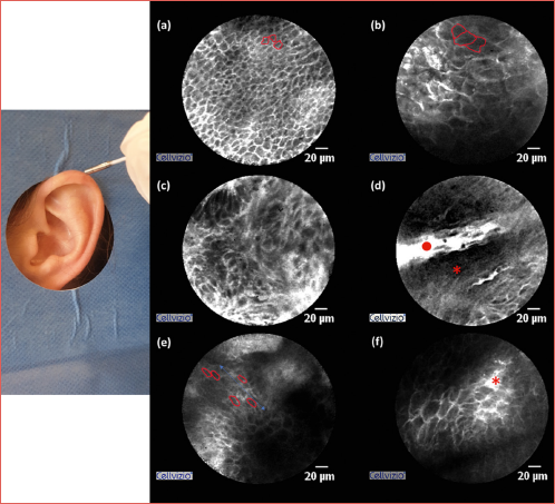
The BCCs in the region of the auricle could all be reached without any problems. The probe was held like a pen in the examiner’s hand and guided over the auricle (Cover figure). In this way, all tumour margins and the surrounding macroscopically healthy tissue could completely be accessed with the CLE probe. Even BCCs that were supposedly more difficult to access, with extension in the concha, the scapha or in the direction of the auditory canal, could be reached effortlessly.
Diagnostic accuracy
In the blinded evaluation of the sequences, all 4 examiners showed high reliability in differentiating between BCC and healthy tissue. The 4 different examiners achieved 81.3-98.4% accuracy, sensitivities of 78.1-90.6%, and specificities of 84.4-100%. The positive predictive values were 83.3-100%, and the negative predictive value were 79.4-97%. This resulted in an average accuracy, sensitivity and specificity of 90.6 ± 6.1%, 86.7 ± 7.4% and 94.5 ± 6.3%. The averaged positive and negative predictive values were 94.2 ± 6.8% and 87.8 ± 6.6%.
Interrater agreement
Fleiss’ kappa was calculated to assess interrater agreement among the 4 investigators. According to Landis and Koch, a κ value of 0.76 was obtained, indicating substantial agreement13.
Assessment of the a priori malignancy criteria
In addition to the evaluation of the sequences by the 4 investigators, one investigator (F.M.) carried out the separate analysis of the 6 a priori criteria on all 64 sequences. This showed that the sequences of BCC met significantly more of the criteria than the sequences with healthy tissue (4 ± 0.8 vs 0.4 ± 0.5, p < 0.001). In addition, each BCC sequence fulfilled at least 3 criteria, while the sequences with healthy tissue met a maximum of 2 criteria per sequence.
If only the individual criteria are taken into account, the criteria inhomogenous tissue, unsharp cell margins, enlarged cells, epidermal shadowing, palisading/polarisation and fibrosis achieved an accuracy of 87.5%, 95.3%, 92.2%, 68.8%, 59.4% and 78.1%. For a further overview of the individual criteria see Figure 1. For a further comparison, the criteria were grouped into 2 over groups based on either CLE data on other tumour entities or RCM data on BCC. For the sequences showing tumour, significantly more criteria based on CLE data were met than criteria based on RCM data. On average, the CLE-based criteria scored 2.8 ± 0.2 out of 3 compared to 1.2 ± 0.7 out of 3 for the RCM-based criteria (p < 0.001).
Tissue characteristics
Overall, the healthy tissue presents with homogeneous, small cells. The cells are sharply demarcated and have approximately the same size. In contrast, the images of BCC are characterised by a distinct inhomogeneity. The cells on the BCC sequences vary in size and are poorly demarcated. The deformed, enlarged cells are a characteristic feature of BCC (Cover figure). Between the cell formations, there are cell-free sections with fibrosis-like areas. In certain sequences, the tissue of the BCC is interrupted by areas free of fibrosis and cells (epidermal shadowing) and in some regions the cells expand in a certain direction (polarisation).
Discussion
The study presented herein evaluated the feasibility of using CLE as a potential optical biopsy technique for BCC in the auricular region for the first time. With a mean sensitivity of 86.7% and a mean specificity of 94.5%, CLE was demonstrated to be a reliable tool for differentiating between BCC and healthy tissue. As CLE has not yet been evaluated in skin tumours, we compared our results to other tumour entities. Sensitivity and specificity in our study are comparable to CLE data from bladder cancer (90%/72%) 9, oesophageal cancer (67-100%/56-98%) 7, and head and neck cancers, including oral (90.1%/87.4%) and laryngeal lesions (45.5-100%/60-100%) 14,15. Additionally, the interrater agreement was consistent with the application of CLE in other tumour entities. For example, for laryngeal carcinomas, a slightly higher interrater reliability was reported (κ = 0.89), whereas, for oral cavity carcinomas, the reliability was very similar to our findings, with a κ value of 0.77 11,14.
As an optical biopsy method tested here for the first time, CLE competes with the methods already used in BCC. The European guideline recommends RCM and optical coherence tomography (OCT) for routine clinical use to determine tumour entity 2. However, due to the poor data so far, no recommendation has been made regarding their use to determine resection margins in BCC 2. Regarding sensitivity and specificity in distinguishing BCC from healthy tissue, CLE shows satisfactory results compared to RCM and OCT. The OCT achieves a comparable sensitivity of 79-94% and a specificity of 85-96% in the detection of BCC, but shows a considerably poorer interobserver agreement of κ = 0.52 17. Another meta-analysis reported that the sensitivity and specificity for OCT was 86.4% and the sensitivity of RCM 100%, but with a poorer specificity of 72.5% 18. The combined use of OCT and RCM also yielded a comparable sensitivity of 82.6% and a specificity of 93.8% 19.
A potential limitation of CLE, in comparison to RCM and OCT, lies in its relatively reduced focal depth and narrower field of view. Specifically, CLE achieves a focal depth of up to 70 μm and a field of view of 325 μm, whereas RCM reaches depths of up to 300 μm and offers a field of view of approximately 1 mm 6,20,21. Consequently, CLE provides less distinct visualisation of higher-order tissue architecture, such as cellular alignment (polarisation). This limitation was evident in our evaluation of malignancy criteria, where complex tissue features, deduced from RCM studies on BCC, were observed significantly less frequently compared to cellular-level characteristics, such as cell size and heterogeneity in CLE tumour sequences. In light of these findings, distinguishing BCC from other tumour types may prove more challenging with CLE than with RCM or OCT. However, this notion remains speculative and warrants confirmation through systematic investigations. Notably, this limitation does not appear to compromise diagnostic sensitivity when differentiating BCC from healthy tissue – provided that appropriate malignancy criteria are applied.
An advantage of CLE over the other 2 modalities (OCT and RCM) lies in the handling of the probe. A reported limitation of both OCT and RCM is their reduced ability to image tumours located in anatomically difficult-to-access areas; the auricle is explicitly mentioned as an example in this context 19. In contrast, the study with CLE presented here showed no difficulties reaching the entire extent of the respective tumour with the small probe. Even locations that were difficult to reach, such as BCC in the region of the scapha or tragus, presented no problems.
Compared to the very high oncological safety of Mohs micrographic surgery, which achieves 5-year cure rates of up to 99%, CLE – based on the average sensitivity observed in this study – does not consistently achieve results comparable to this current gold standard 5. An advantage of CLE over Mohs micrographic surgery is that the surgeon can directly examine tissue in vivo. Since no tissue preparation or transport is required, there is a significant time advantage. However, in order to translate this benefit into clinical routine, future studies must focus on further improving the oncological safety of CLE. A key starting point would be to address one of the main limitations of the present study.
This limitation lies in the absence of standardised, objective assessment criteria among the 4 investigators – such as the implementation of a structured scoring system – which may have contributed to interobserver variability and limited the reproducibility of the findings. Previous studies on CLE examinations of other tumour entities have shown that the introduction of a scoring system is an important approach to improve interrater agreement, especially to improve the results of inexperienced examiners 22,23. In order to create the basis for such a scoring system, various malignancy criteria were examined in the current study. All the malignancy criteria used proved to be promising as they occurred significantly more frequently in the sequences with BCC than in those of healthy tissue. Taking all criteria into account, a cut-off value for a possible score was also obtained, in that the BCC sequences all fulfilled at least 3 of 6 criteria, while the sequences from healthy tissue fulfilled a maximum of 2 criteria. Future studies should aim to formulate a fixed scoring system based on the insights gained here and evaluate it across additional sequences.
Another approach to improving interrater reliability could be objectifying malignancy criteria through image processing software measurements. Other studies have, for example, assessed tissue homogeneity and cell count in this way. Using automated image analysis, Sievert et al. achieved a sensitivity and specificity of 88% and 71.9% in detecting pharyngeal and laryngeal carcinomas based only on the criterion of cell density 10. By measuring tissue homogeneity based on signal intensity at different image locations, a sensitivity and specificity of 81.8% and 86.2% were achieved 24. In addition, using AI-based programmes could offer a further opportunity to objectify the results and provide a method of making the CLE technique accessible to less experienced examiners. Furthermore, future studies should aim to validate these findings in larger patient cohorts. The study presented here is an initial feasibility study.
Nevertheless, this feasibility study successfully generated initial data on the application of CLE in BCC and, through the evaluation of defined assessment criteria, laid the groundwork for a more objective diagnostic approach in the future. Finally, these results encourage us to extend CLE to other skin tumours.
Conclusions
The poor macroscopic delineation of tumour margins in the resection of BCC on the one hand and the considerable aesthetic consequences of resections on the auricle on the other make the use of an optical biopsy in this area highly desirable. The study presented here shows that CLE is a promising method for optical biopsy. Further studies are needed to improve the objectivity of BCC detection, e.g., by introducing a standardised evaluation system and using image processing software or artificial intelligence, and to confirm the promising results in larger patient cohorts.
Conflict of interest statement
The authors declare no conflicts of interest.
Funding
This research was funded by the German Research Foundation DFG, Project: 545049923.
Author contributions
FM, MG, SH: conceptualization; FM, MG: methodology; NP, MA: software; NP, MA, MS, BP: validation; MS, AS; BP, MG, FM: formal analysis; investigation, F.M., M.G., T.M.; resources, M.G., S.H.; FM, MG, MS: data curation; FM, MG: writing – original draft prepa-ration; MS, TM, MS, AS, LA, BP: writing – review and editing; LA, FM: visualization; SH, MG: supervision; project administration; MG: funding acquisition. All authors have read and agreed to the published version of the manuscript.
Ethical consideration
This study was approved by the Institutional Ethics Committee on human research of the Julius-Maximilians-University Würzburg (approval number 154/23_mpz-sc).
The research was conducted ethically, with all study procedures being performed in accordance with the requirements of the World Medical Association’s Declaration of Helsinki.
Written informed consent was obtained from each patient for study participation and data publication.
History
Received: May 31, 2025
Accepted: August 4, 2025
Figures and tables
Figure 1. Proportion of sequences that fulfill the respective malignancy criterion, sorted by histology. For all criteria, there is a significantly more frequent occurrence in the BCC sequences than in the healthy tissue.
| Malignancy criteria based on CLE in other tumour entities |
| Inhomogeneous tissue 22 |
| Non-differentiable cell borders 22 |
| Enlarged cells 10 |
| Malignancy criteria based on RCM in BCC |
| Presence of epidermal shadowing 20 |
| Arrangement of the cells following the same axis and perpendicular to the periphery of the tumour islands (polarisation and palisading)20,25 |
| Fibrosis-like areas surrounding individual cell nests 26 |
| Patient | Age | Gender | Largest tumour extension | Localisation |
|---|---|---|---|---|
| 1 | 58 | M | 1.1 cm | posterior surface |
| 2 | 84 | F | 0.5 cm | helix |
| 3 | 87 | M | 1.7 cm | scapha |
| 4 | 79 | M | 0.8 cm | lobulus |
| 5 | 59 | F | 0.9 cm | tragus |
| 6 | 61 | M | 0.9 cm | posterior surface |
| 7 | 87 | M | 2.9 cm | helix |
| 8 | 82 | M | 1.6 cm | posterior surface |
References
- Cameron M, Lee E, Hibler B. Basal cell carcinoma: epidemiology, pathophysiology, clinical and histological subtypes, and disease associations. J Am Acad Dermatol. 2019;80:303-317. doi:https://doi.org/10.1016/j.jaad.2018.03.060
- Peris K, Fargnoli M, Kaufmann R. EADO”A, EDF”B, ESTRO”C, UEMS”D and EADV”E. European consensus-based interdisciplinary guideline for diagnosis and treatment of basal cell carcinoma-update 2023. Eur J Cancer. 2023;192. doi:https://doi.org/10.1016/j.ejca.2023.113254
- Scrivener Y, Grosshans E, Cribier B. Variations of basal cell carcinomas according to gender, age, location and histopathological subtype. Br J Dermatol. 2002;147:41-47. doi:https://doi.org/10.1046/j.1365-2133.2002.04804.x
- Daviti M, Lallas K, Dimitriadis C. Real-life data on the management of incompletely excised basal cell carcinoma. Dermatology. 2023;239:429-435. doi:https://doi.org/10.1159/000529367
- Tolkachjov S, Brodland D, Coldiron B. Understanding Mohs micrographic surgery: a review and practical guide for the nondermatologist. Mayo Clin Proc. 2017;92:1261-1271. doi:https://doi.org/10.1016/j.mayocp.2017.04.009
- Pilonis N, Januszewicz W, di Pietro M. Confocal laser endomicroscopy in gastro-intestinal endoscopy: technical aspects and clinical applications. Transl Gastroenterol Hepatol. 2022;7. doi:https://doi.org/10.21037/tgh.2020.04.02
- Vaculová J, Kroupa R, Kala Z. The use of confocal laser endomicroscopy in diagnosing barrett’s esophagus and esophageal adenocarcinoma. Diagnostics (Basel). 2022;12. doi:https://doi.org/10.3390/diagnostics12071616
- Martínek J, Kollár M, Krajčíová J. Confocal laser endomicroscopy in diagnosing indeterminate biliary strictures and pancreatic lesions: a prospective pilot study. Rozhl Chir. 2020;99:258-265. doi:https://doi.org/10.33699/PIS.2020.99.6.258-265
- Wu J, Wang Y, Luo W. Diagnostic performance of confocal laser endomicroscopy for the detection of bladder cancer: systematic review and meta-analysis. Urol Int. 2020;104:523-532. doi:https://doi.org/10.1159/000508417
- Sievert M, Aubreville M, Eckstein M. Cellular density and variability in laryngeal and pharyngeal squamous cell carcinoma using confocal laser endomicroscopy. Eur Rev Med Pharmacol Sci. 2023;27:3622-3630. doi:https://doi.org/10.26355/eurrev_202304_32146
- Goncalves M, Aubreville M, Mueller S. Probe-based confocal laser endomicroscopy in detecting malignant lesions of vocal folds. Acta Otorhinolaryngol Ital. 2019;39:389-395. doi:https://doi.org/10.14639/0392-100X-2121
- Becker V, von Delius S, Bajbouj M. Intravenous application of fluorescein for confocal laser scanning microscopy: evaluation of contrast dynamics and image quality with increasing injection-to-imaging time. Gastrointest Endosc. 2008;68:319-323. doi:https://doi.org/10.1016/j.gie.2008.01.033
- Landis J, Koch G. The measurement of observer agreement for categorical data. Biometrics. 1977;33:159-174.
- Sievert M, Oetter N, Mantsopoulos K. Systematic classification of confocal laser endomicroscopy for the diagnosis of oral cavity carcinoma. Oral Oncol. 2022;132. doi:https://doi.org/10.1016/j.oraloncology.2022.105978
- Goncalves M, Iro H, Dittberner A. Value of confocal laser endomicroscopy in the diagnosis of vocal cord lesions. Eur Rev Med Pharmacol Sci. 2017;21:3990-3997.
- Chang T, Liu J, Hsiao S. Interobserver agreement of confocal laser endomicroscopy for bladder cancer. J Endourol. 2013;27:598-603. doi:https://doi.org/10.1089/end.2012.0549
- Cheng H, Guitera P. Systematic review of optical coherence tomography usage in the diagnosis and management of basal cell carcinoma. Br J Dermatol. 2015;173:1371-1380. doi:https://doi.org/10.1111/bjd.14042
- Guida S, Alma A, Shaniko K. Non-melanoma skin cancer clearance after medical treatment detected with noninvasive skin imaging: a systematic review and meta-analysis. Cancers (Basel). 2022;14. doi:https://doi.org/10.3390/cancers14122836
- Aleissa S, Navarrete-Dechent C, Cordova M. Presurgical evaluation of basal cell carcinoma using combined reflectance confocal microscopy-optical coherence tomography: a prospective study. J Am Acad Dermatol. 2020;82:962-968. doi:https://doi.org/10.1016/j.jaad.2019.10.028
- González S, Sánchez V, González-Rodríguez A. Confocal microscopy patterns in nonmelanoma skin cancer and clinical applications. Actas Dermosifiliogr. 2014;105:446-458. doi:https://doi.org/10.1016/j.ad.2013.03.010
- Kose K, Cordova M, Duffy M. Video-mosaicing of reflectance confocal images for examination of extended areas of skin in vivo. Br J Dermatol. 2014;171:1239-1241. doi:https://doi.org/10.1111/bjd.13050
- Sievert M, Mantsopoulos K, Mueller S. Systematic interpretation of confocal laser endomicroscopy: larynx and pharynx confocal imaging score. Acta Otorhinolaryngol Ital. 2022;42:26-33. doi:https://doi.org/10.14639/0392-100X-N1643
- Oetter N, Knipfer C, Rohde M. Development and validation of a classification and scoring system for the diagnosis of oral squamous cell carcinomas through confocal laser endomicroscopy. J Transl Med. 2016;14. doi:https://doi.org/10.1186/s12967-016-0919-4
- Sievert M, Aubreville M, Gostian A. Validity of tissue homogeneity in confocal laser endomicroscopy on the diagnosis of laryngeal and hypopharyngeal squamous cell carcinoma. Eur Arch Otorhinolaryngol. 2022;279:4147-4156. doi:https://doi.org/10.1007/s00405-022-07304-y
- Navarrete-Dechent C, DeRosa A, Longo C. Reflectance confocal microscopy terminology glossary for nonmelanocytic skin lesions: a systematic review. J Am Acad Dermatol. 2019;80:1414-1427.e3. doi:https://doi.org/10.1016/j.jaad.2018.12.007
- Peppelman M, Wolberink E, Blokx W. In vivo diagnosis of basal cell carcinoma subtype by reflectance confocal microscopy. Dermatology. 2013;227:255-262. doi:https://doi.org/10.1159/000354762
Downloads
License

This work is licensed under a Creative Commons Attribution-NonCommercial-NoDerivatives 4.0 International License.
Copyright
Copyright (c) 2026 Società Italiana di Otorinolaringoiatria e chirurgia cervico facciale
How to Cite
- Abstract viewed - 299 times
- PDF downloaded - 84 times