


Skull base surgery
Vol. 46: Issue 4 - August 2026
Estimating nasoseptal flap length for sellar reconstruction: a surgical and radiological study
Authors
Fabio Portella Gazmenga
 , Marcelo Hamilton Sampaio
, Thiago Luis Infanger Serrano
, Emerson Taro Inoue Sakuma
, Mateus Dal Fabbro
, Eulalia Sakano
, Marcelo Hamilton Sampaio
, Thiago Luis Infanger Serrano
, Emerson Taro Inoue Sakuma
, Mateus Dal Fabbro
, Eulalia Sakano
Summary
Cover Image
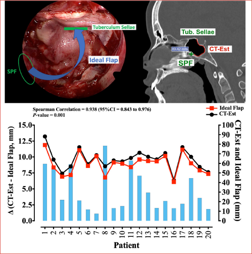
Objective. To assess whether computed tomography (CT) can predict the nasoseptal flap length necessary for reconstruction after endoscopic approaches to the sellar region.
Methods. Twenty consecutive patients who underwent endoscopic resection of pituitary adenoma were included, and data were obtained prospectively in 2 stages, one radiological and one surgical. The length of the flap as estimated by CT (CT-Est) was compared with the flap length considered optimal (Ideal Flap), after intraoperative measurements. Correlation between the 2 values was assessed with the Spearman correlation test.
Results. The mean length estimated on CT scans was 63.8 ± 2.3 mm, and the mean Ideal Flap was 59.1 ± 2.3 mm. The mean difference (Δ CT-Est – Ideal Flap) between these 2 values was 4.7 ± 0.7 mm. The Spearman coefficient of correlation between Ideal Flap and CT-Est was 0.938 (95% CI = 0.843 to 0.976), with p = 0.001, denoting a very strong and statistically significant correlation.
Conclusions. CT-based estimates closely approximated the nasoseptal flap length required for reconstruction of a sellar approach defect. CT has the potential to be a useful tool for preoperative planning of tailored flaps.
Introduction
The advent of the nasoseptal flap (NSF), described by Hadad and Bassagasteguy in 2006 1, revolutionised endoscopic endonasal surgery of the anterior skull base, drastically reducing the rates of postoperative cerebrospinal fluid (CSF) leak and neurological complications 1,2. Radioanatomic studies have demonstrated that the NSF has potential dimensions to cover most anterior skull base defects 3,4. However, in standard sellar approaches for pituitary surgery, the opening at the skull base is so small that the NSF is often excessively large for the proposed reconstruction.
In an attempt to perform less invasive surgery with greater mucosal preservation, research began to focus on the possibility of raising smaller NSFs 5. By sparing the anterior mucosa of the nasal septum and not leaving the septal cartilage exposed, there is less crusting and cartilage injury. This also obviates the need for manoeuvres to cover areas that would otherwise be left bare, such as the use of a reverse flap and mucosal grafts, which have the potential to cause additional sinonasal morbidity 5-7.
However, creating a smaller, custom-made flap remains a challenge due to the concern that it will be insufficient for the proposed reconstruction. The sphenoid sinus has great anatomical variability, and as the flap needs to be in contact with the bone, the extent of pneumatisation directly interferes with the required length of the flap. Likewise, the dimensions of the nasal septum can vary depending on age, sex, and race 8-11.
Therefore, for a smaller NSF to be fashioned safely, it must be tailored to the anatomy of the patient, as informed by preoperative studies. Some radioanatomic studies 3,4,10,11 have estimated the possible dimensions of an NSF and its sufficiency to repair different skull base defects. However, none has tested or applied such estimates in surgical reconstructions. To date, there is no computed tomography (CT)-only model for estimating the size of a flap needed for reconstruction, nor data on whether such a method would be accurate.
Within this context, the objective of this study is to assess whether CT can predict the nasoseptal flap length necessary for reconstruction after endoscopic approaches to the sellar region.
Materials and methods
In this prospective study, data were obtained in 2 stages, one radiological and one surgical, as described below.
Selection of participants
Twenty consecutive patients who underwent endoscopic resection of pituitary adenoma with transsphenoidal access to the sella and a nasoseptal flap raised at the beginning of surgery were included. As a routine in our service, the NSF is raised at the beginning of surgery when an intraoperative CSF leak is expected. Extended approaches, prior radiation therapy, and revision cases were excluded from the sample.
Radiological stage
TECHNICAL ASPECTS
Of the 20 CT scans of the face included, 12 were obtained in a 2008 Toshiba Aquilion TSX-101-A® 64-slice scanner with a 0.5-mm slice protocol. The other 8 scans were obtained in a 2013 GE Healthcare Brightspeed Elite® 16-slice scanner, with a 0.625-mm slice protocol. Multiplanar reconstructions were analysed in Arya 22.12.0® software, certified by the National Health Surveillance Agency.
DEFINITION OF MEASUREMENTS
The sphenopalatine foramen (SPF) was used as the main landmark. It was identified and labeled with a tool that allows its anatomical location to be projected and identified in all CT slices (Fig. 1A). From this projection, 2 tomographic measurements are taken:
- CT-Est. The distance between the projection of the SPF and the tuberculum sellae is measured on the midline sagittal slice. This tortous line drawn over the internal contours of the sphenoid sinus represents the substrate where the NSF is placed during reconstruction. Its measurement is the estimated minimum length that the NSF needs to be in order to cover the defect created by a standard sellar approach (Fig. 1B). This model is similar to that used by Pinheiro-Neto 4 and subsequently reproduced in several other radiological studies. For our purposes, we adapted it to estimate the length needed for reconstruction of an exclusively sellar approach.
- Pedic. In the axial plane, the length of the right flap pedicle (Pedic) is estimated as suggested by Dam et al. 7: after identifying the SPF, 2 perpendicular lines are drawn, one parallel to the medial wall of the maxillary sinus and the other to the posterior wall of the maxillary sinus (Fig. 1C). The distance between the intersection of these lines and the nasal septum corresponds to the length of the flap pedicle (Fig. 1D). Although the pedicle is a curved structure, instead of measuring it directly we chose to measure its projection in a straight line. This method, in addition to being easier and more reproducible, is supported by the results of Dam et al. 7, who demonstrated excellent correspondence between estimated measurement of the pedicle projection and its anatomical dissection.
Surgical stage
SURGICAL TECHNIQUE
In all cases included, the NSF was raised at the beginning of the procedure, and its length (anterior extent) was decided by the surgical team as done routinely. Depending on the anatomy of the sphenoid sinus, tumour, and dimensions of the sella, different flap sizes may be chosen. For all cases, NSF were raised on the right side by the same surgeon.
After decongesting the nasal cavity with epinephrine solution, the inferior and middle turbinates were lateralised. The lower third of the superior turbinate was resected, exposing the natural ostium of the sphenoid sinus, which was then enlarged. With the aid of the monopolar cautery, the NSF was fashioned and positioned in the nasopharynx. The bony posterior septum was detached from the sphenoidal rostrum and all septations within the sphenoid sinus were removed with the aid of low-profile high-speed pneumatic drill. The bony floor is then drilled out. Once the sella has been opened and the tumour resected, a multilayered reconstruction was performed with different materials, such as fat, fascia, or synthetics, as appropriate. The NSF was then positioned so that its entire surface composed of perichondrium and periosteum is in contact with the bone of the sphenoid sinus. The flap must cover the entire sellar defect and any adjacent bone.
INTRAOPERATIVE MEASUREMENTS
Two intraoperative measurements were obtained: SepFlap and Excess (detailed below). Both were performed with the aid of a sterile disposable ruler or a sterilised rigid stainless-steel ruler (Fig. 2A).
All procedures and measurements were performed by the same surgeon.
SEPTAL PORTION OF THE FLAP (SEPFLAP)
After making the incisions that delimit the NSF, the midpoint of the anterior vertical incision is defined as the first landmark (Pant). The second landmark is the most posterior part of the nasal septum, at the level of the arch of the choana. The measurement between these 2 landmarks represents the length of the septal portion of the flap (Fig. 2B).
EXCESS
Once the NSF is in place at the end of the procedure, checks are done to ensure it is in contact with the entire bony surface of the floor of the sphenoid sinus, the clival recess, and the bone adjacent to the sellar defect. Its anterior edge should be as parallel as possible to the tuberculum sellae. The Pant landmark is then identified, and its distance to the tuberculum sellae (Excess) is measured.
This measurement corresponds to how much tissue was left over after the flap reached the tuberculum sellae, i.e., how much was excess tissue beyond that needed to cover the sellar access defect (Fig. 2C-D).
Correlation of CT and surgical data
CALCULATING THE IDEAL FLAP LENGTH
The Ideal Flap corresponds to the flap length that is necessary for the most anterior point of the flap to reach the tuberculum sellae. Its calculation is based on the two measurements obtained intraoperatively and one of the measurements obtained on CT.
The Ideal Flap is calculated as the total dimension of the NSF (its septal portion plus the pedicle) minus any excess tissue: (Ideal Flap = SepFlap + Pedic – Excess) (Fig. 3A-C).
The flap pedicle is an integral part of its total length but is technically difficult to measure intraoperatively due to its posterior location, small dimensions, and curvilinear trajectory. As demonstrated by Dam et al. 7, there is excellent correspondence between the estimated pedicle length measured on CT and its actual length as determined by cadaveric dissection. Therefore, due to the technical difficulty of measuring the pedicle length intraoperatively, its excellent correspondence with tomographic measurements, and little variability in its dimensions, we considered the CT measurement alone for our purposes.
COMPARING CT-EST VERSUS IDEAL FLAP
The length of the flap as estimated by CT (CT-Est) was compared with the Ideal Flap length to determine whether CT can predict the flap size necessary in each case. Both variables are measured between the same two anatomic landmarks, from the sphenopalatine foramen to the tuberculum sellae, one obtained exclusively by preoperative CT (CT-Est) and the other after intraoperative measurements (Ideal Flap) (Fig. 3C-D).
The difference between these variables was defined as CT-Est – Ideal Flap.
Statistical analysis
Categorical data are presented as absolute (N) and relative (%) frequencies. Numerical data are presented as mean ± standard deviation; median (95% confidence interval for the median). Correlation between the 2 methods was assessed with the Spearman correlation test.
Depending on the correlation coefficient (CC) obtained, correlations were classified qualitatively as follows: no correlation (CC = 0); weak (CC = ± 0.001 to ± 0.299); fair or moderate (CC = ± 0.300 to ± 0.599); strong (± 0.600 to ± 0.899); very strong (± 0.900 to 0.999); and full or perfect correlation (CC = 1.00).
Descriptive and inferential statistical analyses were carried out using IBM SPSS Statistics for Macintosh, Version 28.0. Graphical representations were plotted in GraphPad Prism, Version 8.0.0 for Macintosh (GraphPad Software, Boston, Massachusetts, USA; www.graphpad.com). An alpha error of 0.05 was adopted for all statistical analyses.
Results
Patient characteristics
Table I provides a detailed description of the study participants.
CT and intraoperative measurements
Detailed measurements are given in Table II for the CT parameters (CT-Est and Pedic) and intraoperative measurements (SepFlap and Excess).
Correlation of CT and intraoperative data
The mean difference between the length estimated on CT scans (CT-Est) and the Ideal Flap length was 4.7 ± 0.7 mm. Cover figure shows Ideal Flap, CT-Est, and Difference (CT-Est – Ideal Flap) values for each of the 20 cases included. The Spearman coefficient of correlation between Ideal Flap and CT-Est was 0.938 (95% CI = 0.843 to 0.976), with p = 0.001, denoting a very strong and statistically significant correlation.
Discussion
This study evaluated CT as a method to estimate NSF length for sellar reconstruction after endoscopic resection of pituitary adenoma. Our sample of 20 patients had a high incidence of intraoperative CSF leak. This can be explained by the fact that only cases in which the flap was fashioned at the start of surgery, which is usually done when a high likelihood of intraoperative CSF leak is expected, were included in the sample, including large macroadenomas with suprasellar extension and marked stretching and thinning of the diaphragma sellae. Of these cases, 2 exhibited CSF leaks in the postoperative period, but in neither case was the flap considered short or insufficient. It is important to highlight that in many cases the NSF is not harvested at the start of surgery. The “rescue flap” technique allows preservation of the vascular pedicle, with flap elevation performed at the end of the procedure only if an intraoperative CSF leak is identified 12. It is the preferred technique in most skull base centres in cases where a low risk of CSF leak is anticipated 13. In this context, the decision to include only cases in which the flap was harvested at the beginning of surgery was based on methodological considerations. Performing the SepFlap measurement is more appropriate at the beginning of the procedure, when the bony structures are still preserved, in contrast to a rescue flap scenario, where removal of portions of the sphenoid rostrum and the bony nasal septum would compromise the reproducibility and accuracy of the measurements.
Comparison of the CT-based estimate of the flap length (CT-Est) vs the flap length that is considered optimal (Ideal Flap) showed a very strong and statistically significant correlation between these 2 values. CT-Est was greater than Flap-Ideal in all cases; in other words, CT overestimated the flap length (by 4.7 mm on average). This overestimation can be explained by several factors. As the septal portion of the flap is measured with a rigid ruler at the time of surgery, small curves in the mucosa are not taken into account, as suggested by Peris-Celda 14. Likewise, nasal septal deviations in the anteroposterior direction can also contribute to overestimation of the septal part of the flap, as illustrated in Figure 4A.
Finally, the CT estimate is based on anatomy that will be modified by the surgery itself. Bone drilling, especially at the sella, changes the position in which the flap will be supported at the end of the surgery, causing it to diverge from the initially estimated position. Drilling out the sellar bone in cases where the tumour expands into the sphenoid sinus shortens the distance the flap needs to cover to reach the tuberculum sellae (Fig. 4B-E), which is an additional factor in overestimation. Figures 4B and C illustrate the case with the largest difference (CT-Est – Ideal Flap) in this study, highlighting this scenario. On the other hand, in cases with a small or shallow sella, removing the bone may leave the predicted flap position essentially unchanged. Figures 4D and E present the case with the smallest difference (CT-Est – Ideal Flap) in this study, illustrating this situation. Another peculiar scenario is when the clival recess is long in the anteroposterior direction but narrow in the craniocaudal direction. In these cases, the thickness of the flap may prevent it from totally reaching the clival bone as originally predicted on CT scans. Some of these situations may be unpredictable preoperatively and may at least partially explain the discrepancies between the CT-Est and Ideal Flap measurements. Previous radiological studies have discussed the hypothesis that the NSF may exhibit some degree of tissue retraction after it has been harvested and positioned. Pinheiro-Neto et al. 4 consider this possible, while Dam et al. 7 note that, in their clinical experience, this is not relevant. There is no proof or any objective measurement of such possible retraction in the literature. In the 20 cases studied herein, the aforementioned overestimation of flap length seemed to override any immediate retraction that may have occurred. However, subsequent retraction during the healing process is a possibility, as is known to occur in other anatomical regions.
Although CT-based estimates showed a strong correlation with intraoperative measurements, considerable variability was observed among individual cases, as illustrated in Figure 4. In some patients, CT provided a highly precise prediction, whereas in others, a discrepancy of a few millimetres was present, which inside the nasal cavity may be relevant. This observation carries potential practical implications. Previous purely radiologic studies often gave the impression that CT-based estimates would always be accurate, since the measurements relied on well-defined bony landmarks, which made them relatively easy to obtain and replicate. However, as discussed above, our findings demonstrate that while CT estimates can approximate the ideal length, its accuracy may be influenced by some factors, particularly when the bony anatomy is significantly modified during surgery. This observation does not diminish the value of CT as a preoperative planning tool. On the contrary, these findings highlight important nuances that should be incorporated into the design of future studies and the development of standardised preoperative planning protocols.
It bears stressing that this is a preliminary study and that the CT model used to estimate flap length was only compared with the Ideal Flap measurement; it was never tested surgically to inform the creation of a tailored flap. Future studies are needed to test the safety and applicability of a CT-based protocol for creating tailored flaps. In any proposed model, planning a tight flap would be inappropriate, and the addition of a safety margin would be essential for three reasons: first, the possibility of errors in measuring or harvesting the flap, loss of tissue due to use of the cautery, laceration of the flap, and other factors that could lead to insufficiency; second, the possibility of tissue retraction during the healing process; third, as the success of the NSF is dependent on its edges remaining in contact with the bone surrounding the defect, the flap must necessarily be larger than the defect it is meant to cover. It is important to emphasise that the rationale behind using a tailored flap for sellar defect reconstruction is greater mucosal preservation with potentially lower morbidity, but the primary goal and outcome should always be prevention of CSF leak. Raising an insufficient flap is much more harmful than simply missing out on the benefits of a smaller flap.
This is the first study to evaluate the ability of CT to estimate the dimensions of a nasoseptal flap for sellar reconstruction – a possibility that has been extensively studied on CT imaging, but to date had not been compared with intraoperative findings. Our results demonstrated that CT has the potential to be a useful tool in the preoperative planning of smaller nasoseptal flaps, although further clinical validation is necessary. We also encourage further research into alternative tools for this purpose, such as the development of software that would allow 3D reconstruction of estimated measurements, surgical simulation, and 3D printing of models for case-by-case assessment and planning, as recently proposed by Kayastha et al. 6.
This study has some limitations. Given the great variability of sphenoid sinus pneumatisation, our sample of 20 participants may not have encompassed all possible anatomical variants. Although the sample size is limited, this study should be interpreted as a proof-of-concept investigation that may serve as a foundation for future studies with larger cohorts. We also evaluated NSFs only when positioned in a specific location, through the clival recess, while in practice it can be positioned as is most convenient in each case – obliquely or along the lateral wall of the sphenoid, for instance. Finally, another limitation of CT is that, even when using tools that allow 3-dimensional structure localisation and navigation, measurements are always obtained in a 2-dimensional plane, and cannot reflect the truly 3-dimensional surgical procedure.
Conclusions
CT-based estimates closely approximated the nasoseptal flap length required for reconstruction of a sellar approach defect. This study highlights the potential of CT as a planning tool for tailored flaps, though further clinical validation is required to confirm its applicability in surgical practice.
Conflict of interest statement
The authors declare no conflict of interest.
Funding
This research did not receive any specific grant from funding agencies in the public, commercial, or not-for-profit sectors.
Author contributions
FPG: conception, design, data collection, analysis and interpretation, literature review, writing; MHS: conception, design, analysis and interpretation, critical review; TLIS: data collection, analysis and interpretation; ETIS: design, analysis and interpretation; MDF: data collection, analysis and interpretation; ES: supervision, critical review.
Ethical consideration
This study was approved by the Institutional Ethics Committee of the University of Campinas Clinics Hospital, with protocol number 29653420.4.0000.5404. The research was conducted ethically, with all study procedures being performed in accordance with the requirements of the World Medical Association’s Declaration of Helsinki. Written informed consent was obtained from each participant/patient for study participation and data publication.
History
Received: September 8, 2025
Accepted: June 9, 2026
Figures and tables
Figure 1. A) Identification and annotation of the sphenopalatine foramen (red cross) in a coronal CT slice and visualisation of its projection in axial and sagittal slices; B) CT-Est: measurement of the projection of the sphenopalatine foramen (green cross) to the tuberculum sellae in a midline sagittal slice; C) Pedic: 2 perpendicular lines are drawn, one parallel to the medial wall of the maxillary sinus (green) and the other to the posterior wall of the maxillary sinus (yellow); D) The distance between the intersection of these lines and the nasal septum corresponds to the length of the flap pedicle.
Figure 2. A) Sterile rulers used for intraoperative measurements; B) Illustration of the SepFlap measurement obtained intraoperatively: sterile ruler supported on the arch of the choana, measuring from the most anterior point of the nasal septum to the midpoint of the anterior incision (Pant); C) Flap in place, showing the position of the tuberculum sellae (green line), the midpoint of the flap incision (Pant, green asterisk), and the distance between them (Excess, purple double-headed arrow); D) Intraoperative measurement of Excess.
Figure 3. A) Nasoseptal flap in place at the end of surgery. Highlighted in green are the anatomical landmarks used to obtain the measurements of interest; B) Calculation of the Ideal Flap measure (SepFlap – Excess + Pedic); C-D) Representation of the 2 compared measures: Ideal Flap (C) CT-Est (D). Both represent measurements from the sphenopalatine foramen (SPF) to the tuberculum sellae.
Figure 4. A) Nasal septal deviation in the anteroposterior direction. In red, comparison of two methods for measurement of the nasal septum, one in a straight line and the other following its curves; B and D) Preoperative image showing CT-Est, i.e., the estimated minimum size of the nasoseptal flap to cover the defect created by a sellar approach; C and E) Postoperative image showing the possible position of the flap. Removal of the sellar bone shortens the distance required for the flap to reach the tuberculum sellae.
| Characteristic | Group | Data distribution |
|---|---|---|
| Sex* | Female | 9 (45%) |
| Male | 11 (55%) | |
| Intraoperative CSF leak* | No | 8 (40%) |
| Yes | 12 (60%) | |
| Postoperative CSF leak* | No | 18 (90%) |
| Yes | 2 (10%) | |
| Age (years)** | 50.8 ± 4.1; 54.5 (44.5-63) | |
| *Categorical data are presented as absolute (N) and relative (%) frequencies; **Numerical data are presented as mean ± standard deviation; median (95% confidence interval for the median). | ||
| Measurement | Data distribution (mm) |
|---|---|
| SepFlap | 59 ± 2.4; 58.5 (54-66.5) |
| Excess | 12.3 ± 1.8; 11.5 (8-15) |
| CT-Est | 63.8 ± 2.3; 64.1 (58-67.7) |
| Pedic | 12.3 ± 0.2; 12.4 (12.1-12.7) |
| Ideal Flap | 59.1 ± 2.3; 59.9 (54.2-63.2) |
| Difference (Δ CT-Est – Ideal Flap) | 4.7 ± 0.7; 3.2 (2.1-6.8) |
| Numerical data are presented as mean ± standard deviation; median (95% confidence interval for the median). | |
References
- Hadad G, Bassagasteguy L, Carrau R. A novel reconstructive technique after endoscopic expanded endonasal approaches: vascular pedicle nasoseptal flap. Laryngoscope. 2006;116:1882-1886. doi:https://doi.org/10.1097/01.MLG.0000234933.37779.E4
- Cavallo L, Somma T, Solari D. Endoscopic endonasal transsphenoidal surgery: history and evolution. World Neurosurg. 2019;127:686-694. doi:https://doi.org/10.1016/J.WNEU.2019.03.048
- Pinheiro-Neto C, Ramos H, Peris-Celda M. Study of the nasoseptal flap for endoscopic anterior cranial base reconstruction. Laryngoscope. 2011;121:2514-2520. doi:https://doi.org/10.1002/LARY.22353
- Pinheiro-Neto C, Prevedello D, Carrau R. Improving the design of the pedicled nasoseptal flap for skull base reconstruction: a radioanatomic study. Laryngoscope. 2007;117:1560-1569. doi:https://doi.org/10.1097/MLG.0B013E31806DB514
- Barger J, Siow M, Kader M. The posterior nasoseptal flap: a novel technique for closure after endoscopic transsphenoidal resection of pituitary adenomas. Surg Neurol Int. 2018;9. doi:https://doi.org/10.4103/SNI.SNI_192_17
- Kayastha D, Wiznia D, Manes R. 3D printing for virtual surgical planning of nasoseptal flap skull-base reconstruction: a proof-of-concept study. Int Forum Allergy Rhinol. 2023;13:2073-2075. doi:https://doi.org/10.1002/ALR.23165
- ten Dam E, Korsten-Meijer A, Schepers R. Calculating nasoseptal flap dimensions: a cadaveric study using cone beam computed tomography. Eur Arch Otorhinolaryngol. 2015;272:2371-2379. doi:https://doi.org/10.1007/S00405-014-3353-3
- Jeong C, Cho J, Park Y. Differences in the predicted nasoseptal flap length among races: a propensity score matching analysis. PLoS One. 2023;18. doi:https://doi.org/10.1371/JOURNAL.PONE.0283140
- Ramos H, Pinheiro-Neto C, Possatti L. Pneumatization of the sphenoidal sinus may affect endonasal cranial base reconstruction. J Craniofac Surg. 2022;33:E808-E810. doi:https://doi.org/10.1097/SCS.0000000000008690
- Shah R, Surowitz J, Patel M. Endoscopic pedicled nasoseptal flap reconstruction for pediatric skull base defects. Laryngoscope. 2009;119:1067-1075. doi:https://doi.org/10.1002/LARY.20216
- Park S, Kim H, Kim D. Radioanatomic study of the skull base and septum in Asians: implications for using the nasoseptal flap for anterior skull-base reconstruction. Int Forum Allergy Rhinol. 2017;7:999-1005. doi:https://doi.org/10.1002/ALR.21993
- Rivera-Serrano C, Snyderman C, Gardner P. Nasoseptal “rescue” flap: a novel modification of the nasoseptal flap technique for pituitary surgery. Laryngoscope. 2011;121:990-993. doi:https://doi.org/10.1002/LARY.21419
- Gazmenga F, Prevedello D, Carrau R. Skull base reconstruction following endoscopic pituitary surgery: a global survey. J Neurol Surg B Skull Base. 2026;87:299-307. doi:https://doi.org/10.1055/A-2607-1028
- Peris-Celda M, Pinheiro-Neto C, Funaki T. The extended nasoseptal flap for skull base reconstruction of the clival region: an anatomical and radiological study. J Neurol Surg B Skull Base. 2013;74:369-385. doi:https://doi.org/10.1055/S-0033-1347368
Downloads
License

This work is licensed under a Creative Commons Attribution-NonCommercial-NoDerivatives 4.0 International License.
Copyright
Copyright (c) 2026 Società Italiana di Otorinolaringoiatria e chirurgia cervico facciale
How to Cite
- Abstract viewed - 300 times
- PDF downloaded - 78 times