Vestibology
Vol. 46: Issue 3 - June 2026
Side correlation between subjective visual vertical tilt and symptoms in migraine with vertigo
Summary
Cover Image
Objective. To analyse the subjective visual vertical (SVV) in migraine patients with balance disorders and its correlation with symptoms. We hypothesise that vestibular imbalance reflects a global migraine activation leading to symptom prevalence on one side.
Methods. Twenty-six migraine patients with vertigo and/or dizziness (Group A) were studied in the interictal period. Symptoms assessed: unilateral (U) vs bilateral (B) headache (H), cranial autonomic (CAS) or cochlear symptoms (CS), vertigo/dizziness. SVV test was performed in all participants. Group A was compared to Group B (migraine without vertigo/dizziness) and Group C (controls). Main parameters: SVV0 (mean tilt in 6 trials) and SVV side exclusivity (SE: tilt of same sign in ≥ 5 trials).
Results. SVV0A (0.80 ± 0.68) was significantly greater than SVV0B (0.39 ± 0.40; p = 0.01) and SVV0C (0.32 ± 0.19; p = 0.001). SVV SE was found in 78.6% of patients with vertigo and 18.7% with dizziness (p = 0.003). SVV SE incidence was higher in UH (64.7%) vs BH (11.1%; p = 0.02), and in UCAS (72.7%) and UCS (85.7%) vs BCAS/none (26.6%) and BCS/none (31.6%) (p = 0.02; p = 0.04).
Conclusions. A slight SVV tilt persists between vertigo attacks and correlates with migraine imbalance underlying side-prevalent symptoms.
Introduction
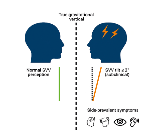
Visual verticality perception is based on a gravitational input that is processed by the central nervous system and whose constitution involves visual, vestibular and proprioceptive signals 1. Determination of Subjective Visual Vertical (SVV) is the main test proposed to assess perception of verticality in both research and clinical practice. In the SVV test, patients must align a visible luminous line, in darkness, with the perfect vertical (marked by gravity) without other visual references. The tilt of SVV is a highly sensitive sign of vestibular tone imbalance in the roll plane 2. If a healthy subject is placed in a dark room, with the only visible object being a phosphorescent line, he is able to place the line in a perfectly vertical position with a margin of error of ± 2° 3. In the presence of a lesion of the peripheral and/or central vestibular pathways, particularly the otolithic ones, the line indicated by the patient as perfectly vertical may not overlap with the true gravitational vertical but may be inclined towards one side 4. The alteration of SVV is always associated with a postural synkinesis known as ocular tilt reaction (OTR) as an expression of a lesion of the central or peripheral otolithic pathways, in particular utricular. The only exception is represented by thalamo-cortical lesions in which the alteration of the SVV can be present in an isolated manner without OTR 5. Among the 3 components that constitute the OTR, skew deviation, head tilt and ocular counter roll, SVV deviation can be considered an indirect manifestation of the latter 2. SVV has been studied in several peripheral vestibular disorders. In acute unilateral vestibulopathy (AUV) the highest values of alteration are recorded and SVV tilt is always directed towards the side of the lesion 6. In benign paroxysmal positional vertigo (BPPV) the amount of the error in determining SVV is always lower than in the AUV and the tilt is variable, being able to be either ipsilesional or contralesional 7,8. Some authors report changes or an immediate improvement in SVV and disequilibrium following canalith repositioning manoeuvre that predicts resolution of BPPV 9. SVV deviation occurred in Meniere’s attacks in a considerable number of patients. The tilt was toward the affected ear and returned to normal within a few weeks after crisis in most cases 10. During acute attacks of Ménière’s disease (MD), unilateral otolithic involvement expressed by SVV tilt may have an opposite trend compared to concomitant canal signs in the early stages of the disease 11. A perceptual error due to an imbalance in the otolithic pathway can also be expected in migraine patients, particularly in those with balance disorders. More generally, it is conceivable that an imbalance in the pathogenetic mechanisms at work can favour the onset of lateral symptoms in migraine. Headache, for example, can more often be unilateral in location but not infrequently, in approximately 40% of cases, bilateral 12. Similarly, cranial autonomic symptoms (CAS) which often accompany migraine attacks can be expressed bilaterally or, on the contrary, exclusively or predominantly on one side 13. On the vestibular side, the imbalance in the pathogenetic mechanisms of migraine translates into symptoms characterized by attacks of spontaneous vertigo typical of vestibular migraine (VM) and similar to MD or in triggered forms such as visual induced vertigo. If the same pathogenetic mechanisms act in a diffuse or bilateral manner, the prevailing symptom will be dizziness 14. Cochlear symptoms (CS), such as fullness, can also be expressed bilaterally or unilaterally, configuring, in the absence of vestibular symptoms, a cochlear migraine 15 or representing the beginning of MD which is the main labyrinthine comorbidity of migraine. The aim of this study was first to verify, through the determination of the SVV, the presence of a vestibular tone imbalance (vestibular asymmetry) in the otolithic pathway in subjects affected by migraine with or without VM or more generally with or without balance disorders such as vertigo or dizziness. Subsequently, an attempt was made to establish a possible correlation between the alteration detected in the determination of SVV and migraine symptoms including headache and CAS skull localisation, the CS and the type of balance disorder (vertigo or dizziness). The authors hypothesise that an imbalance in the vestibular pathway involved in the perception of the SVV reflects a global migraine activation leading to a prevalence of the symptoms on one side. A visual summary of the study concept is provided in the Cover figure, which illustrates SVV tilt relative to the true vertical and its relation with side-prevalent symptoms in migraine.
Materials and methods
Twenty-six consecutive patients (17 females, 9 males; age, 39 ± 11.7) suffering from migraine with episodic, isolated or persistent balance disorders, who came to our observation in the period from 30 January 2022 to 30 December 2024, were recruited. All patients were studied in the interictal period between headache and/or vertigo attacks. The time between the last attack and the evaluation was 8±4.8 days. A thorough medical history was obtained to determine the migraine-like nature of the headache and some associated clinical manifestations. First, the localisation of the headache during attacks was considered. Based on this, the headache was defined as unilateral or predominantly unilateral (unilateral headache: UH) in case of exclusive or prevalent localisation in one half of the skull, or bilateral (BH) with widespread localisation throughout the skull without laterality. Secondly, the type of balance disorder reported by patients was investigated according to the International Classification of Vestibular symptoms, which distinguishes 2 categories: vertigo (spontaneous or triggered) and dizziness (spontaneous or triggered) 16. In each patient, it was also verified whether the diagnostic criteria for VM (according to the Bárány Society) were met 17. The possible coexistence of headache with CAS including rhinorrhoea, nasal obstruction, lacrimation and conjunctival hyperaemia was also evaluated 13. In this study, the patient is considered to be affected by CAS in the presence of at least one of the above-mentioned symptoms during attack. As for headache, the predominant or exclusive unilaterality or bilaterality of the symptoms was considered. Finally, auricular fullness, hearing loss and tinnitus, not necessarily associated with one other or with the headache attack, and their localisation (unilateral or bilateral) were considered among the CS. All patients underwent neurological and neuroradiological evaluation (brain MRI) to exclude ischemic, degenerative, demyelinating or expansive lesions of the brain. The same neurological evaluation was used to define primary migraine headache in doubtful cases. In addition, audiological and vestibular tests were performed to exclude patients with vestibular disorders unrelated to migraine. Vestibular function was assessed through the following tests: positional, caloric, cervical vestibular evoked myogenic potentials (cVEMPs), vestibular head impulse test (vHIT) as well as the SVV test. From a statistical point of view, the prevalence of the clinical parameters identified through the medical history in the group of patients examined (Group A) was calculated. SVV data emerging from Group A were compared with those of two homogeneous groups matched for gender and age: Group B consisting of 26 participants affected by migraine without balance disorders (i.e., vertigo and/or dizziness) and Group C consisting of 26 normal subjects not affected by migraine and without balance disorders. The comparison between percentages of the clinical parameters under examination was carried out using the chi square test with and without Yates correction. The level of statistical significance is set for values of p < 0.05.
SVV Test
For the test, we used a 30-cm-high fluorescent bar with LED, which was placed in a darkened room and set 1m away from the subject. The bar was mounted on the wall via central rod, around which the bar could be rotated manually or via remote control in both directions (right or left). At the upper end, there was a pointer that slides with the bar on a graduated scale, in which 0° degree corresponds to perfect alignment of the longitudinal line of the bar with the direction of gravity. Rotation of the pointer to the left corresponds to negative angles expressed in degrees with a maximum resolution of 0.5°, whereas rotation to the right corresponds to positive angles. The test involved the use of a dynamic image composed of a pointed illuminated sight that could slide along the longitudinal axis of the bar, with a pendular motion at a frequency of 36 cycles/min (dynamic test) 3. The test was performed with the patient standing upright holding his/her head in straight position with a soft foam support (40x40 cm) placed between the patient’s feet and the floor (provocative test)18. In this condition, 6 measurements were conducted, alternating the bar from a 45° angle to the right and then to the left, for a total of 3 measurements per side. The mean value of the 6 measurements (SVV0), preceded by the + or – sign was the primary study parameter for each individual subject. The mean value of the individual SVV0, without the + or – sign (absolute value), was the main parameter referred to each group (SVV0A, SVV0B, SVV0C) under examination. The distribution of the SVV values of the 6 measurements with reference to the 0° value was also taken into consideration. In particular, our attention was directed to those who presented a side exclusivity in the distribution of the SVV values (SVV side exclusivity: SVV SE). With this definition we have identified those whose values of the 6 measurements were all of the same sign (- or +) or, at most, 5 of the same sign and 0° in the other one (Tab. I).
This limit was established because in a group of normal subjects, matched for number, gender and age (Group C of the study), the best result obtained in the distribution of the individual 6 values on one side was 4 of the same sign, one of the opposite, and the last one without any sign, respectively (see subjects 7 and 20 of Group C). The need to define a SVV SE as a study parameter lies in the possibility of identifying a subclinical perceptual imbalance, where the SVV tilt values in a subject are contained within +/- 2°, this considered as the normal range 6.
SVV SE was furthermore compared to the skull location (left or right) of the UH, UCAS and UCS in order to verify the presence of a side correlation.
The comparison between means and standard deviations of the SVV perceptual errors was carried out using the t-test or alternatively, when normality in the distribution of the two samples was not satisfied, the non-parametric Mann-Whitney test.
Results
In all patients in Group A, the SVV0 measurement – reflecting individual error in verticality perception – was always within ± 2°, which is considered the normal range6. Overall, patients in Group A showed a mean of the absolute SVV0 values (SVV0A) of 0.8 ± 0.68. For Group B and Group C, SVV0B and SVV0C were 0.39 ± 0.4 and 0.32 ± 0.19, respectively. Thus, a statistically significant increase in perceptual error in determining SVV was observed in Group A compared with both Group B (p = 0.01) and Group C (p = 0.001). No significant difference (p = 0.40) emerged between the 2 control groups (B vs C). An SVV SE was found in 46.1% of Group A patients, in 15.2% of Group B, and in 3.8% of Group C. Comparison between Group A and Group B revealed a statistically significant increase in SVV SE incidence in Group A (χ2 = 4.2824; p = 0.038). The difference was even more significant when comparing Group A to Group C (χ2 = 12.4103; p = 0.004) and remained significant with Yates’ correction (χ2 = 10.2564; p = 0.001). No significant difference was found between Group B and Group C (p = 0.08) (Tab. II).
Among the balance disorders considered, vertigo was reported in 14 of the 26 patients in Group A (53.8%). In all cases, it was episodic and never isolated. Dizziness was reported in 16 patients (61.5%): in 12 cases as the only symptom, and in 4 together with vertigo. Dizziness was episodic in 7 cases, isolated in 4, and persistent in 5. Of the 14 patients with a history of vertigo, 11 (78.6%) presented SVV SE. In contrast, only 3 of the 16 patients with dizziness (18.7%) showed the same. Thus, SVV SE incidence was significantly higher (p = 0.003) in patients with vertigo (χ2 with Yates’ correction = 8.4668). The diagnostic criteria for VM were met in 9 of 26 patients (34.6%) in Group A. All reported episodic vertigo, and 2 also reported dizziness. SVV SE was present in 8 of the 9 patients with VM (88.8%), and in 4 of the 17 without VM (21%). The difference was statistically significant (p = 0.005; χ2 with Yates’ correction = 7.6562). In 17 Group A patients, UH was exclusively or predominantly present in most migraine attacks. The remaining 9 had BH. SVV SE was found in 11 of the 17 patients with UH (64.7%) and in one of the 9 with BH (11.1%). The incidence was significantly higher in the UH subgroup (p = 0.02; χ2 with Yates’ correction = 4.8158). CAS were reported by 13 of 26 patients in Group A (50%). In 11 cases (42.3%), they were unilateral in at least one migraine attack; in 2 cases (7.7%), they were bilateral or diffuse. Among those with unilateral CAS, 9 patients (81.8%) also had UH and 8 (72.7%) experienced vertigo. SVV SE occurred in 8 of the 11 with unilateral CAS (72.7%) and in 4 of the remaining 15 (26.6%). This difference was statistically significant (p = 0.02; χ2 = 5.4176, without Yates’ correction). CS, including both unilateral and bilateral forms, were reported in 16 patients (61.5%) in Group A and 8 (31%) in Group B. This difference was statistically significant (p = 0.026; χ2 = 4.9524). Unilateral CS were found in 7 Group A patients (26.9%) and in one Group B subject (3.8%), again a significant difference (p = 0.021; χ2 = 5.3182). A SVV SE concerned 6 (85.7%) of the 7 Group A patients with unilateral CS and 6 (31.6%) of the remaining 19 (bilateral CS and no CS patients). This difference was statistically significant both without (p = 0.01; χ2 = 4.0506) and with Yates’ correction (p = 0.04) (Tab. III).
All vestibular tests were normal in Group A patients and no statistically significant differences were found in terms of value and asymmetry ratio with the control Groups B and C. Side correlation between SVV SE and symptoms related to migraine are showed in Table IV.
Discussion
Several methods have been proposed for studying SVV. A reliable, though expensive and complex, method involves the study of the otolithic reflex in the OFF-ON vertical axis condition 19. The most common methods are the bucket test 7 and the assessment of SVV in a darkened environment 3. In this case, the visual reference generally consists of a light bar that slides along a frontal plane and is fixed against a background that is either stationary and unlit (static test) or illuminated and moving (dynamic test). The method used to assess SVV in this study, as described in our previous work 3,18, involves a dynamic and provocative test. This method increases the test’s sensitivity compared to that performed under static conditions, especially some time after the acute episode 3. It is therefore particularly useful in cases of vestibular dysfunction such as migraine since, unlike in unilateral vestibular loss, the alterations affecting the otolithic system are expected to be milder and transient, with a tendency toward functional recovery without any lasting effect.
The luminous moving reference we used, consisting of a pointed illuminated target swinging along the longitudinal axis of the light bar, demands greater effort in fixation and smooth pursuit compared to a static reference. The increased engagement of visuo-oculomotor reflexes toward the spatial context of interest enhances the relevance of visual references within the bar while reducing attention to external visual cues. This operates as a sort of functional concentric narrowing of the visual field 6. Such a strategy is valuable because achieving a perfectly dark room to eliminate visual reference cues has always been a limitation in SVV assessment methods.
Furthermore, the use of foam placed between the subject’s feet and the floor (provocative test) reduces the compensatory influence of extra-vestibular sensory input. The importance of exteroceptive and proprioceptive signals from the plantar surface in constructing vertical perception has been demonstrated, especially in the context of vestibular dysfunction18.
Data in the literature regarding SVV in patients with migraine – with or without vestibular symptoms, particularly VM – are quite mixed. Some authors report no significant SVV alterations in VM patients and suggest that the bucket test helps differentiate VM from other central vestibular disorders, in which SVV is often tilted 20. Other authors found no difference in verticality misperception prevalence among patients with primary headaches 21. Indeed, the mean absolute error in degrees was not significantly different between patients with migraine, tension-type headache, and healthy controls.
However, a systematic review and meta-analysis including 7 studies and 816 participants showed misperception of visual verticality in patients with migraine and tension-type headache compared to controls when assessed via the SVV test 22. Vestibular migraine subjects did not show significant deviation in static SVV but did demonstrate greater deviation in dynamic tasks 23. This finding was confirmed by later studies, where alterations were also detected using dynamic subjective visual horizontal testing. Both measures were significantly more affected than the static test in VM subjects compared to controls 24.
The evaluation of migraine patients in our study was always conducted in the interictal period, outside of headache or vertigo episodes. Therefore, the SVV error measured through 6 consecutive trials always remained within normal limits. These results are expected, considering that vestibular function, although transiently impaired during attacks, likely returns to normal between episodes. Our findings thus differ from the aforementioned studies, where the timing of test execution relative to symptom onset was not specified. It is possible that in those studies the tests were conducted closer to symptomatic episodes, potentially explaining the greater SVV deviations reported. In fact, in our study, the average time between evaluation and the last episode was 8 ± 4.8 days.
Nevertheless, our study reveals subclinical misperception in patients belonging to Group A, particularly those meeting VM criteria 14. This alteration, reflected by increased SVV0 values, was not observed in Groups B or C. This becomes more relevant when considering SVV SE, which was also significantly higher in Group A patients. Otolithic imbalance appears to correlate with the type of balance disorder reported. Higher SVV0 and SVV SE values were more prevalent in patients with a history of episodic vertigo. This may reflect dysfunction in migraine-related mechanisms acting on vestibular centres.
It is well established that various neuropeptides and neurotransmitters involved in migraine pathophysiology, such as 5-hydroxytryptamine, noradrenaline, dopamine, and calcitonin gene-related peptide, can modulate vestibular neuron sensitivity. Unilateral release of these substances may cause vestibular system imbalance and vertigo, while bilateral release may result in hypersensitivity and dizziness without clear vestibular asymmetry 14. Similarly, UH might correspond to unilateral release, and in general, the localisation of migraine-related symptoms (including CAS and cutaneous allodynia) may depend on how these substances are released.
Our study shows that misperception of verticality correlates significantly with the imbalance in migraine-related pathophysiological mechanisms. SVV assessment may serve not only as a reliable indicator of vertigo but also of all unilateral symptoms manifested in migraine. Although a significant correlation emerged between perceptual imbalance and migraine symptoms in patients with balance disorders, especially episodic vertigo, no strict correlation was found between the direction of SVV tilt and the predominant side of symptoms. This might reflect the current functional state of the vestibular otolithic pathway on the side most affected by migraine activation. In cases of hypofunction, SVV tilt might be expected toward the symptomatic side; the opposite may occur in hyperfunction.
Animal electrophysiological and neuroanatomical studies, along with human neuroimaging (PET, fMRI), have identified the ventroposterolateral and ventroposteromedial thalamic nuclei and the vestibular cortex as components of the central vestibular system 25. The thalamus is involved in multisensory integration and processing of vestibular, visual, and proprioceptive inputs. Thalamic activation has been demonstrated during migraine attacks via fMRI 25. Based on these connections, it is plausible that migraine attacks transiently sensitise thalamo-cortical pathways, enhancing sensory perception and spatial orientation 26. The perception of verticality, an advanced cognitive function, can be considered part of this multimodal integration.
For these reasons, the SVV test can reveal vestibular tone imbalance in the roll plane caused by dysfunction anywhere from the labyrinthine receptors to the cortex, even in the absence of OTR 5. SVV is therefore the most frequently altered test in otolithic system involvement. Furthermore, the presence of an OTR–SVV dissociation (i.e., absent OTR with present SVV tilt) may reflect selective dysmodulation of thalamo-cortical vestibular pathways by migraine mechanisms. This speculation is supported by studies showing that SVV recovery occurs earlier than ocular torsion following acute unilateral vestibular loss 27. In summary, this dissociation may indicate high-level (thalamo-cortical) interaction between migraine and vestibular systems, whereas the simultaneous presence of OTR or a reversed dissociation (OTR present with no SVV tilt) might reflect involvement at a lower (brainstem) level.
The correlation with lateralised migraine symptoms observed in our study may support the hypothesis that verticality misperception may originate in thalamo-cortical pathways involved in global sensory processing. In any case, this finding always requires exclusion of other neurological causes such as infarction 5. The patients in our study showed no functional imbalance in conventional vestibular testing. However, the degree of SVV tilt we recorded was small (subclinical) and detectable only using our enhanced method. Thus, this result has limited value for comparison.
In contrast, a comparative analysis between SVV tilt and ocular torsion, both expressed in degrees, would be desirable during migraine attacks, when greater roll-plane imbalance is expected. A perceptual system analysis focused on tonic responses does not necessarily require ocular VEMPs, as these reflect the function of the transient system not involved in tonic otolithic responses 28.
The significance of a persistent SVV alteration outside of migraine episodes remains unclear. Based on our findings, it may reflect an imbalance that not only affects the vestibular pathway but also involves the broader pathophysiological mechanisms of migraine, ultimately influencing its clinical manifestations. However, it remains uncertain whether this condition, despite apparently normal performance on conventional tests, represents a true vulnerability of the vestibular system with potential long-term clinical implications. Further longitudinal studies are needed to determine whether subclinical SVV alteration between attacks may hold prognostic value.
Conclusions
Our study highlights a persistent imbalance in the perception of verticality in migraine patients with balance disorders, particularly in those experiencing episodic vertigo. The degree of this misperception is mild during the interictal period and becomes more detectable when the SVV test is conducted under the dynamic and provocative conditions we employed, as these enhance the test’s sensitivity. The introduction of SVV SE, obtained through repeated measurements of SVV0, proved to be a useful parameter for increasing the statistical significance of the data under analysis. Verticality misperception correlates significantly with the imbalance of the pathogenic mechanisms broadly involved in migraine. Therefore, SVV tilt serves not only as a reliable indicator of vertigo, but also of all lateralised symptoms associated with the migraine phenotype.
Conflict of interest statement
The authors declare no conflict of interest.
Funding
This research did not receive any specific grant from funding agencies in the public, commercial, or not-for-profit sectors.
Ethical consideration
The study was conducted in accordance with the Declaration of Helsinki and approved by the Ethics Committee of Perugia Hospital (protocol code: DREAM24, register number: 4813/24, date of approval: 16/10/2024).
The research was conducted ethically, with all study procedures being performed in accordance with the requirements of the World Medical Association’s Declaration of Helsinki.
Written informed consent was obtained from each participant/patient for study participation and data publication.
History
Received: September 13, 2025
Accepted: December 18, 2025
Figures and tables
| Subject | 1st test | 2nd test | 3rd test | 4th test | 5th test | 6th test | SVV0 (mean) |
|---|---|---|---|---|---|---|---|
| Right vestibular dysfunction | +3° | +3° | +3° | +3° | +3° | +3° | +3° |
| Left vestibular dysfunction | -4° | -4° | -4° | -4° | -4° | -4° | -4° |
| Normal | 0° | 0° | 0° | 0° | 0° | 0° | 0° |
| Right SVV SE (subclinical dysfunction) | +2° | +2° | +2° | +2° | +2° | +2° | +2° |
| Left SVV SE (subclinical dysfunction) | -2° | -2° | -2° | -2° | -2° | -2° | -2° |
| SVV | A vs B | p | SVV | A vs C | p | SVV | B vs C | p |
|---|---|---|---|---|---|---|---|---|
| SVV 0 A | 0.8 ± 0.68 | 0.01 | SVV0 A | 0.8 ± 0.68 | 0.001 | SVV0 B | 0.39 ± 0.4 | 0.40 |
| SVV 0 B | 0.39 ± 0.4 | SVV0 C | 0.32 ± 0.19 | SVV0 C | 0.32 ± 0.19 | |||
| SVV SE A | 46.1% | 0.038 | SVV SE A | 46.1% | 0.001 | SVV SE B | 15.2% | 0.08 |
| SVV SE B | 15.2% | SVV SE C | 3.8% | SVV SE C | 3.8% | |||
| A: group A; B: group B; C: group C. SVV0 A, B, C: mean absolute values of SVV0 related to group A, B, C. SVV SE: SVV side exclusivity. | ||||||||
| Group A | SVV SE | no SVV SE | p value |
|---|---|---|---|
| VM Criteria fullfilled (9 patients) | 8 (88.9%) | 1 (11.1%) | 0.005 (YC) |
| VM Criteria not fullfilled (17 patients) | 4 (23.5%) | 13 (76.5%) | |
| Vertigo (14 patients) | 11 (78.6%) | 3 (21.4%) | 0.003 (YC) |
| Dizziness (16 patients) | 3 (18.7%) | 13 (81.3%) | |
| Unilateral H (17 patients) | 11 (64.7%) | 6 (35.3%) | 0.02 (YC) |
| Bilateral H (9 patients) | 1 (11.1%) | 8 (88.9%) | |
| Unilateral CAS (11 patients) | 8 (72.7%) | 3 (27.3%) | 0.02 |
| Bilateral CAS and without CAS (15 patients) | 4 (26.7%) | 11 (73.3%) | |
| Unilateral CS (7 patients) | 6 (85.7%) | 1 (14.3%) | 0.01 |
| Bilateral CS and without CS (19 patients) | 6 (31.6%) | 13 (68.4%) | |
| VM: vestibular migraine; H: headache; CAS: cranial autonomic symptoms; CS: cochlear symptoms; YC: yates correction. | |||
| 1 Only patients in Group A who meet the criteria for SVV SE are reported in this table. | SVV SE | UH | UCAS | UCS |
|---|---|---|---|---|
| 2 The only 2 patients who show side correlation between SVV SE and all 4 symptoms are shown in bold. | Left | Right | Right | |
| 5 | Right | Left | Left | Left |
| 9 | Right | Right | ||
| 11 2 | Right | Right | Right | Right |
| 14 | Left | Left | ||
| 15 | Right | Left | Left | Left |
| 17 | Left | Right | Right | |
| 18 | Right | Left | Left | |
| 20 | Right | Right | Right | |
| 22 2 | Right | Right | Right | Right |
| 23 | Left | Left | ||
| 26 | Right | Right | Right | |
| UH: unilateral headache; UCAS: unilateral cranial autonomic symptoms; UCS: unilateral cochlear symptoms. | ||||
References
- Mazibrada G, Tariq S, Perennou D, et al. The peripheral nervous system and the perception of verticality. Gait Posture 2008;27:202-208. https://doi.org/10.1016/j.gaitpost.2007.03.006
- Dieterich M, Brandt T. Ocular torsion and tilt of subjective visual vertical sensitive brainstem signs. Ann Neurotol 1993;33:292-299. https://doi.org/10.1002/ana.410330311
- Faralli M, Ricci G, Molini E, et al. Determining subjective visual vertical: dynamic versus static testing. Otol Neurotol 2007;28:1069-1071. https://doi.org/10.1097/mao.0b013e31815aea1b
- Brandt T, Dieterich M. Pathological eye-head coordination in roll: tonic ocular tilt reaction in mesencephalic and medullary lesions. Brain 1987;110:649-666. https://doi.org/10.1093/brain/110.3.649
- Dieterich M, Brandt T. Perception of verticality and vestibular disorders of balance and falls. Front Neurol 2019;10:1-15. https://doi.org/10.3389/fneur.2019.00172
- Bohmer A, Rickenmann J. The subjective visual vertical as a clinical parameter of vestibular function in peripheral vestibular diseases. J Vestib Res 1995;5:35-45.
- Chetana N, Jayesh R. Subjective visual vertical in various vestibular disorders by using a simple bucket test. Indian J Otolaryngol Head Neck Surg 2015;67:180-184. https://doi.org/10.1007/s12070-014-0760-0
- Haijoub S, Lacour M. Asymmetry of the SVV in patients with unilateral peripheral vestibular deficit. J Audiol Otol 2024;28:213-220. https://doi.org/10.7874/jao.2023.00346
- Little CC, Schwam ZG, Campo M, et al. Immediate improvement in subjective visual vertical and disequilibrium predicts resolution of benign paroxysmal positional vertigo following single canalith repositioning manuever. Otol Neurotol Open 2022;2:E014. https://doi.org/10.1097/ONO.0000000000000014
- Kumagami H, Sainoo Y, Fujiyama D, et al. Subjective visual vertical in acute attacks of Meniere’s disease. Otol Neurotol 2009;30:206-209. https://doi.org/10.1097/MAO.0b013e3181925010
- Faralli M, Lapenna R, Mandalà M, et al. The first attack of Meniere’s disease: a study through SVV perception, clinical and pathogenetic implications. J Vestib Res 2014;24:335-342. https://doi.org/10.3233/VES-140533
- Randolph W, Evans, MD. The clinical features of migraine with and without aura. Pract Neurol 2014;Apr:26-32.
- Karsan N, Nagara k, Goadsby PG. Cranial autonomic symptoms: prevalence, phenotype and laterality in migraine and two potentially new symptoms. J Headache Pain 2022;23:18-25. https://doi.org/10.1186/s10194-022-01389-w
- Lempert T. Vestibular migraine. Semin Neurol 2013;33:212-218. https://doi.org/10.1055/s-0033-1354596
- Tsung LJ. Cochlear migraine: a possible cause of hearing loss and tinnitus. Hearing J 2018;71:8-9. https://doi.org/10.1097/01.HJ.0000547398.36713.6f
- Bisdorff A, Von Brevern M, Lempert T, et al. Classification of vestibular symptoms: towards an international classification of vestibular disorders. J Vestib Res 2009;19:1-13. https://doi.org/10.3233/VES-2009-0343
- Lempert T, Olesen J, Furman J, et al. Vestibular migraine: diagnostic criteria. J Vestib Res 2012;22:167-172. https://doi.org/10.3233/VES-2012-0453
- Faralli M, Longari F, Ricci G, et al. Influence of extero-and proprioceptive afferents of the plantar surface in determining subjective visual vertical in patients with unilateral vestibular dysfunction. Acta Otorhinolaryngol Ital 2009;29:245-250.
- Byun JY, Hong SM, Yeo SG, et al. Role of subjective visual vertical test during eccentric rotation in the recovery phase of vestibular neuritis. Auris Nasus Larynx 2010;37:565-569. https://doi.org/10.1016/j.anl.2010.02.004
- Chang T, Winnick AA, Hsu YC, et al. The bucket test differentiates patients with MRI confirmed brainstem/cerebellar lesions from patients having migraine and dizziness alone. BMC Neurology 2019;19:219-217. https://doi.org/10.1186/s12883-019-1442-z
- Rodríguez-Almagro D, Obrero-Gaitán E, Lomas-Vega E, et al. New mobile device to measure verticality perception: results in young subjects with headaches. Diagnostics 2020;10:796. https://doi.org/10.3390/diagnostics10100796
- Gaitán EO, Navarro MM, Ortega MAL, et al. Misperception of visual verticality in patients with primary headache disorders: a systematic review with meta-analysis. Brain Sci 2020;10:664. https://doi.org/10.3390/brainsci10100664
- Miller MA, Crane BT. Static and dynamic visual vertical perception in subjects with migraine and vestibular migraine. World J Otorhinolaryngol Head Neck Surg 2016;2:175-180. https://doi.org/10.1016/j.wjorl.2016.08.001
- Ashish G, Augustine AM, Tyagi AK, et al. Subjective visual vertical and horizontal in vestibular migraine. J Int Adv Otol 2017;13:254-258. https://doi.org/10.5152/iao.2017.4056
- Lopez C, Blanke O. The thalamocortical vestibular system in animals and humans. Brain Res Ves 2011;67:119-146. https://doi.org/10.1016/j.brainresrev.2010.12.002
- Espinoza-Sanchez JM, Lopez-Escamez JA. New insights into pathophysiology of vestibular migraine. Front Neurol 2015;12:1-6. https://doi.org/10.3389/fneur.2015.00012
- Faralli M, Ricci G, Manzari L, et al. Different time course of compensation of subjective visual vertical and ocular torsion after acute unilateral vestibular lesion. Eur Arch Otorhinolaryngol 2021;278:2263-2276. https://doi.org/10.1007/s00405-020-06312-0
- Cherchi M. Utricular function in vestibular neuritis: a pilot study of concordance/discordance between ocular vestibular evoked myogenic potentials and ocular cycloposition. Exp Brain Res 2019;237:1531-1538. https://doi.org/10.1007/s00221-019-05529-8
Downloads
License

This work is licensed under a Creative Commons Attribution-NonCommercial-NoDerivatives 4.0 International License.
Copyright
Copyright (c) 2026 Società Italiana di Otorinolaringoiatria e chirurgia cervico facciale
How to Cite
- Abstract viewed - 277 times
- PDF downloaded - 75 times