Reviews
Online First
Clinical utility and advances of liquid biopsy in hypopharyngeal carcinoma: a systematic review and meta-analysis
Summary
Cover Image
Background. Hypopharyngeal carcinoma (HPC) is an aggressive malignancy with poor prognosis due to difficult early diagnosis, frequent recurrence, and distant metastasis. Although surgery, radiotherapy, chemotherapy, and immunotherapy have progressed, accurate prognostic assessment and real-time monitoring remain limited. Liquid biopsy, a minimally invasive method detecting circulating tumour DNA (ctDNA), circulating tumour cells (CTCs), and microRNAs (miRNAs), shows promise in solid tumours, but its role in HPC is underexplored.
Methods. A systematic search of PubMed, Embase, and Web of Science was conducted to September 2025 following PRISMA guidelines. Eligible studies evaluated ctDNA, CTCs, or miRNAs for diagnosis, treatment monitoring, or prognosis of HPC, reporting sensitivity, specificity, diagnostic odds ratio, or hazard ratio for meta-analysis.
Results. Twenty-four studies with 933 patients were included. Pooled estimates showed sensitivity 88.3% (95% CI: 84.7-91.6) and specificity 95.9% (95% CI: 93.3-97.9), with predictive intervals sensitivity 0.74–0.98 and specificity 0.85-1.00. Heterogeneity was moderate (I² sensitivity = 52.3%; specificity = 48.5%), and no publication bias was observed. Subgroup analyses indicated ctDNA was advantageous for treatment monitoring, CTCs were consistent in prognosis, and miRNAs were strong candidates due to stability and detection sensitivity. Sensitivity analyses confirmed robustness.
Conclusions. Liquid biopsy shows strong potential for diagnosis, monitoring, and prognosis in HPC. Large-scale, multicentre studies are required to validate its clinical application and integration into patient-management pathways.
Introduction
Hypopharyngeal carcinoma (HPC) 1 is a subtype of head and neck squamous cell carcinoma (HNSCC) that originates in the pyriform sinus, posterior pharyngeal wall, and retrocricoid region. Although it accounts for only 3-5% of all head and neck cancers, it is regarded as one of the most aggressive and lethal malignancies because of its concealed anatomical location, propensity for submucosal infiltration with the majority diagnosed at advanced stages. Hypopharyngeal cancer accounts for 0.3-0.5% of all malignant tumours in China and predominantly occurs in males, with a male-to-female ratio of 5:1 to 10:1, most of whom are diagnosed after the age of 50, while smoking and alcohol consumption remain the major risk factors 2.
The management of HPC requires a multidisciplinary approach, incorporating surgery, radiotherapy, and systemic therapies 3,4. The goal of treatment is to control the tumour while preserving voice and swallowing functions. For patients with T3-T4 category or cartilage invasion, total laryngectomy or wide resection of the hypopharynx and larynx is the standard operation 5. For patients with T1-T2 category disease, organ sparing surgery (e.g. laser microsurgery or robotic surgery) can be selected. Since the rate of cervical lymph node metastasis is as high as 60%, bilateral or selective neck dissection is usually required 6. Radiotherapy is an important component of adjuvant treatment, and intensity-modulated radiotherapy (IMRT) is often recommended. Postoperative radiotherapy is particularly indicated for high-risk pathological factors such as positive surgical margins, extracapsular extension or multiple positive lymph nodes, perineural invasion, and vascular invasion 7,9. For patients who refuse surgery or wish to preserve laryngeal function, cisplatin-based concurrent chemoradiotherapy can be used, but the long-term laryngeal function preservation rate is limited. In recent years, immune checkpoint inhibitors (ICIs), including nivolumab and pembrolizumab, have demonstrated survival benefits in recurrent or metastatic HNSCC, and the Checkmate-141 trial confirmed that nivolumab can improve overall survival in such patients, including the hypopharyngeal tumours 8,9. Although it has not yet become the standard of care in definitive treatment, ICIs are being included in multiple clinical trials. They are being increasingly incorporated into clinical trials for postoperative residual or refractory disease. Despite continuous advances in treatment, the overall prognosis of HPC is still not ideal, especially for patients with locally advanced disease, the 5-year survival rate is less than 50%, and the risk of distant metastasis is high 10. As a potential auxiliary tool, liquid biopsy can enable early detection and real-time monitoring, providing new possibilities for improving treatment effectiveness and patient prognosis.
However, reliable haematological biomarkers to achieve this goal are still lacking. Imaging examinations, such as CT, PET, MRI, and fibreoptic laryngoscopy, remain the main tools for diagnosis and follow-up 11. Squamous cell carcinoma antigen (SCC-Ag) and carcinoembryonic antigen (CEA) have been investigated in hypopharyngeal carcinoma, but their diagnostic and prognostic value is limited 12. Therefore, there is an urgent need to identify novel biomarkers for early diagnosis, prediction of therapeutic efficacy, and monitoring of disease progression.
Liquid biopsy, an emerging molecular diagnostic technique, detects tumour-derived components such as circulating tumour DNA (ctDNA), circulating tumour cells (CTCs), cell-free RNAs (including microRNAs [miRNAs] and various non-coding RNAs), and extracellular vesicles (EVs) in blood, saliva, or other body fluids, enabling non-invasive cancer detection 13-15. Compared with traditional tissue biopsy, liquid biopsy has the advantages of minimally invasive, repeatable sampling and real-time reflection of tumour burden and evolution 16. In a variety of solid tumours, such as lung cancer, breast cancer and colorectal cancer, liquid biopsy has shown important value in early screening, minimal residual disease (MRD) monitoring, recurrence prediction and treatment response evaluation 17. By monitoring ctDNA, CTCs and miRNAs in blood, liquid biopsy can reflect the changes of tumour burden in real time, provide timely and accurate support for clinical decision-making, and potentially improve patient outcomes. In HPC, although research remains at an early stage and is limited in scale, accumulating evidence indicates that ctDNA mutations, aberrant miRNAs expression profiles, and CTCs detection are closely associated with tumour progression, therapeutic response, and patient prognosis.
Nevertheless, most of the existing studies are exploratory with small sample sizes and lack standardised detection methods as well as systematic quantitative analyses. Up to now, there has been no meta-analysis to systematically evaluate the diagnostic value and prognostic significance of liquid biopsy in HPC. Therefore, the aim of this study is to comprehensively evaluate the clinical application value of ctDNA, CTCs and miRNAs in the diagnosis, treatment monitoring and prognostic prediction of HPC through a systematic review and meta-analysis, and to explore their complementarity with traditional imaging and pathological methods.
This study is expected to provide evidence-based support for future related research and the application of liquid biopsy in the individualised management of HPC.
Materials and methods
Literature search strategy
This systematic review and meta-analysis adhered to the 2020 PRISMA guidelines. The protocol was registered with the International Prospective Register of Systematic Reviews (PROSPERO, Registration No. CRD420251153035). A comprehensive literature search was conducted in the PubMed, Embase, and Web of Science databases up to September 2025. The terms used for searching were: “Hypopharyngeal carcinoma” AND “liquid biopsy” AND “circulating tumour DNA” AND “circulating tumour cells (CTCs)” AND “microRNAs (miRNAs)” AND “prognosis” AND “diagnosis” AND “treatment monitoring”. A combination of Boolean operators (AND, OR) was employed to retrieve exhaustive and relevant research articles.
Inclusion and exclusion criteria
Inclusion criteria were: (1) confirmed hypopharyngeal cancer through histopathological examination; (2) use of liquid biopsy technology (e.g., ctDNA, CTCs, miRNAs) for diagnosis, treatment monitoring, or prognostic evaluation 18; (3) provision of sufficient data to extract sensitivity, specificity, diagnostic odds ratio (DOR), or hazard ratio (HR); (4) prospective or retrospective clinical study design; (5) publication in English.
The exclusion criteria were as follows: (1) reviews, case reports, expert opinions or conference abstracts; (2) non-clinical studies, such as animal experiments or in vitro experiments; (3) studies with incomplete data or without extracted outcome indicators; (4) studies with a sample size of less than 10 patients.
Data extraction
Data were independently extracted by 2 researchers using a standardised data collection form. The extracted information included: first author’s name, year of publication, study design (prospective/retrospective), country, sample size, type of liquid biopsy biomarker (ctDNA, CTCs, miRNAs), detection technology (ddPCR, qPCR, NGS, etc.), sensitivity, specificity, and follow-up time and clinically relevant indicators (such as HR, AUC, etc.). In case of missing data or any dispute, the corresponding author was contacted immediately for reconfirmation. We also performed sensitivity analyses to ensure the robustness of the results, and subgroup analyses based on different detection platforms (e.g., NGS, ddPCR, qPCR) and biomarker types (e.g., miRNAs, ctDNA) were performed to further verify the differences in diagnostic performance.
Quality assessment
The methodological quality of eligible studies was evaluated using the QUADAS-2 tool, covering 4 domains: patient selection, detection methods, gold standard and the risk of bias in the reporting of results. Quality assessment was performed independently by 2 researchers. Disagreements were resolved through consensus or adjudication by a third researcher.
Statistical analysis
All statistical analyses were performed using Stata version 17.0 (StataCorp, College Station, TX, USA) and Meta-DiSc version 1.4. A random effects model was used to integrate the sensitivity, specificity, positive likelihood ratio (PLR), negative likelihood ratio (NLR), and DOR, and the 95% confidence interval (95% CI) was calculated 19. The pooled receiver operating characteristic (ROC) curve was drawn and the area under the curve (AUC) was calculated to evaluate the overall diagnostic efficacy. Between-study heterogeneity was assessed by Cochran’s Q test and I2 statistic 20. I2 ≥ 50% indicated significant heterogeneity, so the random effects model was used for effect pooling 21. If I2 was less than 50%, heterogeneity was considered to be acceptable, and a fixed-effect model was used. Subgroup analyses were conducted to explore potential sources of heterogeneity. Publication bias was evaluated using funnel plots. All analyses were conducted using a random effects model given the expected clinical and methodological heterogeneity. To verify the robustness of the results, we conducted multiple sensitivity analyses. The leave-one-out analysis method is used to exclude each study separately and test the impact of each individual study on the overall analysis results. In addition, subgroup analysis (for example, grouping by test platform such as NGS, ddPCR, qPCR and biomarker type such as miRNA, ctDNA) is used to explore differences in diagnostic performance of different tests and biomarkers to ensure the reliability of the results. All analyses were performed using a random effects model, ensuring that the heterogeneity of the data was adequately assessed.
Results
Literature screening
Initially, a total of 518 records were retrieved using database searches, including 121 articles from PubMed, 274 articles from Embase and 123 from Web of Science. After the duplicates were removed, a total of 200 records remained. In addition, 56 articles that were not consistent with the research types (animal experiments, case reports, reviews, guidelines, and expert opinions) were excluded, and after the preliminary screening of titles and abstracts, the primary screening included a total of 144 articles. After full-text screening, 24 eligible studies were finally included that involved 933 patients (Fig. 1).
Characteristics of the studies
The 24 studies included were published between 2015 and 2025 and were from China, Japan, Australia and The Netherlands (Tab. I). The studies were divided into 2 types: half were prospective studies, and half were retrospective studies. All disease stages were covered among studies. Liquid biopsy technology dominated in each study. Digital droplet PCR (ddPCR), real-time quantitative PCR (qPCR) and next generation sequencing (NGS) were the platforms for detection. Positive rates varied from 7.4% to 31.2%, while the negative rates varied from 67% to 93.2%. The distribution of quality scores significantly different, ranging from 1 to 14, which implied that studies differed in methodological quality. The ranges of overall follow-up time spanned from 6 to 48 months.
Although Table I summarises the basic characteristics of the included studies, several noteworthy patterns deserve explicit clarification. First, the positive rate variability (7.4-31.2%) reflects the heterogeneity of detection platforms and biomarker types, with lower positive rates mostly seen in CTC-based studies, whereas miRNA-based studies showed higher positivity. Second, the quality score range (1-14) indicates methodological inconsistency across studies, as lower-quality studies tended to lack standardised cut-off values or prospective design. Third, differences in tumour stage distribution (early vs advanced) may influence diagnostic performance because ctDNA/CTCs are more abundant in late-stage disease. Finally, the studies were predominantly based in China, which may limit external applicability; however, European and Australian cohorts showed similar diagnostic patterns, suggesting generalisability.
Overall diagnostic accuracy
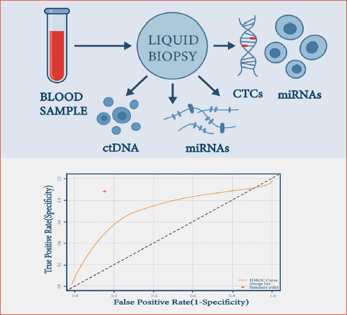
In the meta-analysis, a total of 24 studies involving 933 patients were included. The forest plot showed that the sensitivity and specificity of most studies were more than 0.80, and the results were relatively distributed, suggesting that the overall diagnostic efficacy was high and the consistency was good. The pooled sensitivity of the assay was 0.88 (95% CI: 0.85 to 0.92), with a prediction interval of 0.74 to 0.98 (Fig. 2A), and moderate heterogeneity (I2 = 52.3%, τ2 = 0.0072, p = 0.0016). The pooled specificity was 0.96 (95% CI: 0.93-0.98), the prediction interval was 0.85 to 1.00 (Fig. 2B), and the heterogeneity was moderately low (I2 = 48.5%, τ2 = 0.0067, p = 0.0044). Additional diagnostic performance metrics further supported the strong clinical utility of liquid biopsy in hypopharyngeal carcinoma. The pooled PLR was 21.44 (95% CI: 12.13-37.88), with a clear tendency to significantly increase the probability of the positive result when the result of liquid biopsy is positive. The NLR was 0.12 (95% CI: 0.09-0.16) which is considered important to rule out negative results. DOR was 176.14 (95% CI: 92.83-334.24) indicating an high overall diagnostic efficacy. The ROC curve had an AUC of 0.97 (95% CI: 0.95-0.98), indicating very good discriminatory power (Fig. 2C).
Subgroup analysis
Subgroup analysis was performed in order to explore the potential heterogeneity, stratified by biomarker type and detection platform. There were no significant differences in diagnostic accuracy between ctDNA, CTCs and miRNAs (Tab. II). The pooled sensitivity/specificity/DOR/AUC were 0.89/0.96/176.14/0.97, 0.84/0.95/152.63/0.95 and 0.91/0.97/188.27/0.98, for ctDNA, CTCs and miRNAs, respectively. MiRNAs showed the best diagnostic performance among all the markers, with a sensitivity of 91% and a specificity of 97%. This suggests that miRNA may be the most promising marker in liquid biopsy, especially for early diagnosis and disease surveillance. In contrast, ctDNA and CTCs performed slightly worse, with lower sensitivity and specificity than miRNA. This difference may be related to the dynamic changes of ctDNA and CTCs in tumour progression, especially in the low or early stages of tumour burden.
When stratified by detection method, NGS yielded the best performance (sensitivity 0.92, specificity 0.97), followed by ddPCR (0.88/0.95), whereas qPCR demonstrated relatively lower accuracy (0.85/0.93). These values indicate that a positive liquid biopsy test strongly increases the probability of HPC, whereas a negative test substantially reduces the likelihood of disease. For the detection platform, NGS showed the best sensitivity and specificity among all methods, which may be related to its characteristics of high-throughput and precise detection. ddPCR followed closely and showed high accuracy, especially in the detection of low levels of ctDNA. However, the performance of qPCR was relatively weak and may be affected by the limitations of the detection technology itself.
Detailed subgroup results are listed in Table II.
Sensitivity analyses and robustness assessment
To assess the robustness of the pooled results, multiple sensitivity analyses were performed. Leave-one-out analysis showed that sequential omission of any single study caused only marginal fluctuations in pooled sensitivity, specificity, PLR, NLR, and DOR values, confirming that no individual study disproportionately influenced the overall estimates (Fig. 3A). Subgroup analysis stratified by methodological quality revealed comparable pooled estimates between high-quality (0.939, I2 = 32.4%) and low-quality studies (0.952, I2 = 51.8%), suggesting that part of the heterogeneity may arise from lower-quality designs, yet the overall conclusions remained consistent (Fig. 3B). Study influence analysis further demonstrated that exclusion of individual studies resulted in < 10% variation in I2, with no evidence of outlier effects. Notably, exclusion of Dong et al. yielded an I2 value (45.7%) closest to that of the complete model, indicating that this study was representative of the overall evidence base rather than a heterogeneity-driving outlier (Fig. 3C). For completeness, cumulative meta-analysis and heterogeneity change matrix analyses were also performed, further confirming the stability of effect sizes and the consistency of heterogeneity (Supplementary Figure 1). Collectively, these findings confirm the robustness and reliability of the meta-analytic results.
Publication bias
Publication bias of included studies was checked by funnel plots (Fig. 4), which revealed that points were generally symmetric within the inverted funnel, indicating that no publication bias existed.
Discussion
This systematic meta-analysis revealed that liquid biopsy achieved high diagnostic accuracy, with pooled sensitivity of 0.85-0.92, specificity of 0.93-0.98, a DOR of 176.14, and an AUC of 0.97, outperforming individual studies. Subgroup analyses revealed that biomarker type and detection platform were contributors to interstudy heterogeneity, and higher diagnostic accuracy was seen for miRNAs and NGS-based assays 22,23. The sensitivity analyses did not change the pooled result. Specifically, ctDNA measures the tumour load dynamically and is a good indicator for therapy assessment and disease progression monitoring 24. Because CTC counts are closely related to metastasis and prognosis, they can be potentially used to assess recurrence risk 49,50; and miRNA detection, based on its high stability and sensitivity in body fluid can be regarded as an ideal marker for liquid biopsy applications.
Our research results are consistent with those of previous meta-analyses on head and neck squamous cell carcinoma and solid tumours. This further enhances the reliability of the research results. For instance, in HNSCC, Sun et.al. showed that CTC positivity was significantly associated with both overall survival (OS) and disease-free survival (DFS) 51, while Chen et.al. showed its diagnostic accuracy and association with tumour stage and lymph node metastasis 52. Similar results were reported for lung and breast cancer where ctDNA was highly sensitive for MRD and recurrence as well as survival prediction 53-55. Similarly, for miRNA, diagnostic sensitivity and stability have been verified in gastric and breast cancer, allowing a theoretical foundation for application to HPC. These findings taken together imply that the diagnostic and prognostic utility of liquid biopsy is not HPC-specific but a general biological principle in several malignancies, providing evidence that it is potentially relevant to head and neck cancer in general.
Liquid biopsy has led to novel diagnostic and management possibilities for HPC in the area of personalised medicine by offering a non-invasive, repeatable, and dynamic profiling 56. Liquid biopsy overcomes disadvantages associated with sampling difficulty and tumour heterogeneity that are encountered with histological biopsy. For these reasons, it could be especially valuable for postoperative surveillance and in cases where tissue is not available. Liquid biopsy can be effectively combined with traditional imaging (such as CT, MRI, PET) to make up for the limitations of radiologic examinations in detecting small metastases or small tumours. Traditional imaging usually only provides a macroscopic picture of the tumour, while liquid biopsy can reflect the evolution of the tumour at the molecular level. By combining the two tests, clinicians can obtain more comprehensive patient information, helping to more accurately assess tumour progression, determine treatment response, and predict the risk of recurrence, so as to optimize the patient’s treatment plan. An integrated strategy of liquid biopsy and imaging may function not merely for HPC, but also for other subtypes of HNSCCs.
While our findings are meaningful, there are some limitations. First, there is currently no standardisation for liquid biopsy platforms and the methodological differences could interfere with reproducibility 57. Second, the heterogeneity could be due to the different biomarker selection, thresholds, follow-up time, and definition of endpoints. Third, the studies included are mainly single-centre with a relatively small number of patients and, as a consequence, the subgroup subgroup analyses were underpowered 58. For future studies, standardisation in the detection technique, combination of multimodality of biomarkers and implementation of artificial intelligence in data analysis are recommended. Ultimately we need large, multicentre prospective trials that can confirm the clinical utility in HPC 59,60. Given the shared molecular mechanisms within HNSCC, lessons learned from HPC may accelerate the adoption of liquid biopsy across the head and neck cancer spectrum.
Clinical implications and integration into current therapeutic pathways
Liquid biopsy may complement and potentially improve current clinical workflows for HPC 61.
(1) Early diagnosis: HPC is often diagnosed at advanced stages due to its deep anatomical location and nonspecific symptoms. A blood-based test using miRNA or ctDNA, with pooled sensitivity of 88-91% and specificity of 96–97%, could serve as a pre-endoscopic triage tool, identifying high-risk patients who require urgent laryngoscopy 62.
(2) Treatment monitoring: dynamic changes in ctDNA may indicate treatment response earlier than imaging. For example, a decline in ctDNA levels after induction chemotherapy or concurrent chemoradiotherapy may predict partial/complete response, while persistent positivity may indicate non-response or minimal residual disease 63.
(3) Recurrence surveillance: post-treatment surveillance currently relies on imaging every 3-6 months; however, CT/MRI can miss microscopic recurrence. Liquid biopsy, particularly miRNA and ctDNA assays, can detect molecular relapse. This may allow pre-clinical detection weeks to months earlier than imaging.
(4) Prognostic stratification: CTC positivity is associated with metastasis risk and worse survival. A patient with persistently detectable CTCs after surgery may warrant intensified systemic therapy or closer follow-up intervals 64.
(5) Integration with therapeutic decisions: biomarker-positive patients may benefit from earlier initiation of systemic therapies (chemotherapy or immune checkpoint inhibitors), adaptive treatment modification, or enrolment in biomarker-guided clinical trials.
Conclusions
Our comprehensive review indicates that ctDNA, CTCs and miRNAs display potential clinical significance for the diagnosis, surveillance, and prognostic assessment of HPC. Among these, ctDNA has merits in monitoring disease dynamics and evaluating treatment response; CTCs show strong associations with risk of metastasis and survival outcomes; and miRNAs, owing to their superior stability and sensitivity, are good candidates as clinical biomarkers. Liquid biopsy, as a non-invasive, dynamic and real-time tool, can supplement imaging and histology in enhancing diagnostic accuracy and recurrence surveillance.
Nevertheless, due to the absence of standardised platforms and heterogeneity of the study designs, the present evidence is still not directly comparable and generalised. More future work should be prospective and multicentre with shared consensus of selection of the biomarkers and detection platforms, in terms of reliability. While this study was restricted to HPC, these results can reflect on the wider application of the liquid biopsy in the area of head and neck cancers to develop precision and personalized management. Future work should focus not only on validating diagnostic performance but also on defining standardised clinical algorithms that determine how and when liquid biopsy should be incorporated into HPC management.
Acknowledgements
The authors would like to thank the Department of Head and Neck Surgery, Chaoyang Central Hospital, for their valuable technical support. We also appreciate the assistance of colleagues who provided methodological advice and data extraction validation during the preparation of this systematic review and meta-analysis.
Conflict of interest statement
The authors declare no conflict of interest.
Funding
This work was supported by the Liaoning Natural Science Foundation Program (No. 2019-ZD-0901) and the Liaoning Provincial Natural Science Foundation (No. 2024-MS-290).
Author contributions
LL: study concepts, study design, manuscript editing, manuscript review; JL, YW: data acquisition; LZ, JZ, JL: quality control of data and algorithms; JL: data analysis and interpretation; YW, JL: statistical analysis; JL: manuscript preparation; all authors; final approval of manuscript.
Ethical consideration
This study is a secondary analysis based on previously published literature and publicly available data. Therefore, ethical approval and informed consent were not required. All procedures were conducted in accordance with the ethical standards of the World Medical Association’s Declaration of Helsinki.
Supplementary material
Supplementary figure 1. Cumulative meta-analysis and heterogeneity assessment. (A) Meta-analysis under cumulative order, the pooled effect size converged quickly with being stable as more studies were added and confidence intervals narrowed, indicating the time-consistency of results; (B) Heterogeneity (I2) values recalculated after sequential exclusion of individual studies. No single study markedly altered I2, confirming that overall heterogeneity was not disproportionately driven by any individual study.
History
Received: October 5, 2025
Accepted: December 21, 2025
Figures and tables
Figure 1. PRISMA flow diagram of study selection process. A total of 518 records were identified from PubMed, Embase, and Web of Science. After screening and eligibility assessment, 24 studies were included in the meta-analysis.
Figure 2. Pooled diagnostic performance of liquid biopsy for hypopharyngeal carcinoma. (A) Forest plot of pooled sensitivity across 24 studies. The overall sensitivity was 0.88 (95% CI: 0.85-0.92) with moderate heterogeneity (I2 = 52.3%), and the prediction interval was 0.74-0.98; (B) Forest plot of pooled specificity across 24 studies. The overall specificity was 0.96 (95% CI: 0.93-0.98) with moderate heterogeneity (I2 = 48.5%), and the prediction interval was 0.85-1.00; (C) Hierarchical summary receiver operating characteristic (HSROC) curve summarizing overall diagnostic performance. The red cross indicates the pooled sensitivity (0.88) and specificity (0.96). The area under the curve (AUC) was 0.97 (95% CI: 0.95-0.98), indicating excellent discriminatory power and robust diagnostic accuracy.
Figure 3. Sensitivity analyses for the robustness of the results of the meta-analysis. (A) Leave-one-out sensitivity analysis of pooled positive rate. Each dot represents the pooled estimate after omitting one study, with horizontal lines indicating 95% CIs. The narrow and overlapping intervals confirm that no single study disproportionately influenced the overall estimates; (B) Subgroup analysis by study quality. High-quality studies showed lower heterogeneity (I2 = 32.4%) compared with low-quality studies (I2 = 51.8%), although effect sizes remained consistent across subgroups; (C) Influence analysis illustrating that sequential exclusion of individual studies did not cause > 10% variation in I2, indicating no undue influence on the overall conclusions.
Figure 4. Funnel plot for publication bias assessment. Funnel plot generated using the Freeman-Tukey double arcsine transformed proportion. The plot shows a generally symmetrical distribution of studies around the pooled effect size, suggesting no significant publication bias.
| Study | Country | N | Stage (I-II) (n) | Stage (III-IV) (n) | Diagnostic method | Positive | Negative | Quality assessment score |
|---|---|---|---|---|---|---|---|---|
| Xu, 2019 22 | China | 382 | 188 | 194 | LB | 24.2 | 76.5 | 5 |
| Yang, 2024 23 | China | 20 | 10 | 10 | LB | 7.3 | 92 | 1 |
| Wu, 2021 24 | China | 78 | 40 | 38 | LB | 14.2 | 86.1 | 2 |
| Poel, 2020 25 | Netherlands | 83 | 58 | 25 | LB | 12.8 | 82.6 | 2 |
| Zhang, 2024 26 | China | 17 | 10 | 7 | LB | 26.9 | 70.7 | 6 |
| Wang, 2020 27 | China | 50 | 25 | 25 | LB | 25.3 | 76.5 | 2 |
| Liu, 2023 28 | China | 40 | NS | NS | LB | 31.2 | 69 | 2 |
| Wang, 2018 29 | China | 54 | 12 | 42 | LB | 9 | 89.8 | 1 |
| Zhang, 2016 30 | Australia | 43 | 36 | 17 | LB | 18.3 | 82.6 | 0 |
| Gao, 2022 31 | China | 60 | 30 | 30 | LB | 7.5 | 85.5 | 3 |
| Xu, 2019 32 | China | 30 | NS | NS | LB | 12.7 | 67 | 6 |
| Tian, 2015 33 | China | 76 | 40 | 36 | LB | 20.6 | 92.5 | 13 |
| Kikkawa, 2014 34 | Japan | 46 | 23 | 23 | LB | 7.4 | 88.8 | 4 |
| Hu, 2024 35 | China | 22 | 11 | 11 | LB | 12.1 | 72.2 | 7 |
| Kikkawa, 2010 36 | Japan | 20 | 10 | 10 | LB | 24.5 | 71.3 | 6 |
| Meng, 2022 37 | China | 10 | 5 | 5 | LB | 21.6 | 69.7 | 7 |
| Wu, 2023 38 | China | 117 | 80 | 37 | LB | 12.7 | 74.6 | 3 |
| Feng, 2011 39 | China | 100 | NS | NS | LB | 22.9 | 80.6 | 9 |
| Liu, 2020 40 | China | 91 | 53 | 38 | LB | 28.9 | 78.1 | 6 |
| Fukumoto, 2014 41 | Japan | 44 | 22 | 22 | LB | 6.8 | 74.3 | 1 |
| Lin, 2021 42 | China | 19 | NS | NS | LB | 28.8 | 82.9 | 14 |
| Li, 2022 43 | China | 113 | 51 | 62 | LB | 25.9 | 74.2 | 4 |
| Wei, 2023 44 | China | 56 | NS | NS | LB | 16 | 74.3 | 10 |
| Kinoshita, 2013 45 | Japan | 46 | 20 | 26 | LB | 10.9 | 76.5 | 7 |
| LB, liquid biopsy; NS, not specified. | ||||||||
| Type of biomarker evaluated | No. of studies (n) | Total sample size (n) | Pooled sensitivity (95% CI) | Pooled specificity (95% CI) | DOR (95% CI) | AUC (95% CI) |
|---|---|---|---|---|---|---|
| ctDNA | 10 | 400 | 0.89 (0.85-0.92) | 0.96 (0.93-0.98) | 176.14 (92.83-334.24) | 0.97 (0.95-0.98) |
| CTCs | 8 | 300 | 0.84 (0.78-0.89) | 0.95 (0.91-0.97) | 152.63 (85-270) | 0.95 (0.92-0.97) |
| miRNA | 6 | 233 | 0.91 (0.86-0.94) | 0.97 (0.94-0.99) | 188.27 (100-350) | 0.98 (0.96-0.99) |
| ctDNA: circulating tumour DNA; CTCs: circulating tumour cells; miRNAs: microRNAs; DOR: diagnostic odds ratio; AUC: area under the curve; CI: confidence interval. | ||||||
References
- Wycliffe N, Grover R, Kim P. Hypopharyngeal cancer. Top Magn Reson Imaging. 2007;18:243-258. doi:https://doi.org/10.1097/RMR.0b013e3181570c3f
- Hu J, Yan J, Chen Y. ESCO2 promotes hypopharyngeal carcinoma progression in a STAT1-dependent manner. BMC Cancer. 2023;23. doi:https://doi.org/10.1186/s12885-023-11684-7
- Luo X, Yi J. Progress on multi-disciplinary combined therapy for locally advanced hypopharyngeal carcinoma. Cancer Res Prev Treat. 2023;50:327-333. doi:https://doi.org/10.3971/j.issn.1000-8578.2023.22.1296
- Gui L, Xie Z, Zhang W. Tislelizumab plus nab-paclitaxel and cisplatin as induction immunochemotherapy for organ preservation of locally advanced hypopharyngeal squamous cell carcinoma: a single-arm phase II trial. ESMO Open. 2025;10. doi:https://doi.org/10.1016/j.esmoop.2025.105505
- Hoffmann T. Total laryngectomy – Still cutting-edge?. Cancers (Basel). 2021;13. doi:https://doi.org/10.3390/cancers13061405
- Meulemans J, Delaere P, Vander Poorten V. Primary treatment of T1-T2 hypopharyngeal cancer: changing paradigms. Adv Otorhinolaryngol. 2019;83:54-65. doi:https://doi.org/10.1159/000492310
- Ampil F, Ong C-A, Lian T. Postoperative management in laryngeal cancer with subglottic extension and histologically negative nodes: which patients need adjuvant radiotherapy?. Ear Nose Throat J. 2014;93:354-360. doi:https://doi.org/10.1177/014556131409300814
- Harrington K, Ferris R, Blumenschein G. Nivolumab versus investigator’s-choice single-agent therapy in recurrent or metastatic squamous cell carcinoma of the head and neck (CheckMate 141): health-related quality-of-life results from a randomised, phase 3 trial. Lancet Oncol. 2017;18:1104-1115. doi:https://doi.org/10.1016/S1470-2045(17)30421-7
- Ferris R, Blumenschein G, Fayette J. Nivolumab vs investigator’s choice in recurrent or metastatic squamous cell carcinoma of the head and neck: 2-year survival update of CheckMate 141 with analyses by tumour PD-L1 expression. Oral Oncol. 2018;81:45-51. doi:https://doi.org/10.1016/j.oraloncology.2018.04.008
- Nishimaki T, Kanda T, Nakagawa S. Outcomes and prognostic factors after surgical resection of hypopharyngeal and cervical oesophageal carcinomas. Int Surg. 2002;87:38-44.
- Chan J, Wei W. Current management of hypopharyngeal carcinoma. Auris Nasus Larynx. 2013;40:2-6. doi:https://doi.org/10.1016/j.anl.2011.11.009
- Barak V, Meirovitz A, Leibovici V. Diagnostic and prognostic value of tumour markers (CEA, SCC, CYFRA 21-1, TPS) in head and neck cancer patients. Anticancer Res. 2015;35:5519-5524.
- Nikanjam M, Kato S, Kurzrock R. Liquid biopsy: current technology and clinical applications. J Hematol Oncol. 2022;15. doi:https://doi.org/10.1186/s13045-022-01351-y
- Yu D, Li Y, Wang M. Exosomes as a new frontier of cancer liquid biopsy. Mol Cancer. 2022;21. doi:https://doi.org/10.1186/s12943-022-01509-9
- Li W, Liu J, Hou L. Liquid biopsy in lung cancer: significance in diagnostics, prediction, and treatment monitoring. Mol Cancer. 2022;21. doi:https://doi.org/10.1186/s12943-022-01505-z
- Shegekar T, Vodithala S, Juganavar A. The emerging role of liquid biopsies in revolutionising cancer diagnosis and therapy. Cureus. 2023;15. doi:https://doi.org/10.7759/cureus.43650
- Urbania T, Dusendang J, Herrinton L. Standardized reporting and management of suspicious findings on chest CT is associated with improved lung cancer diagnosis in an observational study. Chest. 2020;158:2211-2220. doi:https://doi.org/10.1016/j.chest.2020.05.595
- Zheng M, Zhou Q, Chen H. Cerebrospinal fluid circulating tumour DNA profiling for risk stratification and matched treatment of CNS metastases. Nat Med. 2025;31:1547-1556. doi:https://doi.org/10.1038/s41591-025-03538-5
- Furuya-Kanamori L, Meletis E, Xu C. Overconfident results with the bivariate random-effects model for meta-analysis of diagnostic accuracy studies. J Evid Based Med. 2022;15:6-9. doi:https://doi.org/10.1111/jebm.12467
- Bowden J, Tierney J, Copas A. Quantifying, displaying and accounting for heterogeneity in meta-analysis of RCTs using standard and generalised Q statistics. BMC Med Res Methodol. 2011;11. doi:https://doi.org/10.1186/1471-2288-11-41
- Spineli L, Pandis N. Statistical heterogeneity: notion and estimation in meta-analysis. Am J Orthod Dentofacial Orthop. 2020;157:856-859.e2. doi:https://doi.org/10.1016/j.ajodo.2020.03.009
- Xu X, Lu Z, Gross N. A 3-miRNA signature predicts survival of patients with hypopharyngeal squamous cell carcinoma after post-operative radiotherapy. J Cell Mol Med. 2019;23:8280-8291. doi:https://doi.org/10.1111/jcmm.14702
- Yang X, Feng C, Jiang D. circ0005027 acting as a ceRNA affects the malignant biological behavior of hypopharyngeal squamous cell carcinoma by modulating miR-548c-3p/CDH1 axis. Biochem Genet. 2024;62:2853-2868. doi:https://doi.org/10.1007/s10528-023-10570-y
- Wu P, Fang X, Liu Y. N6-methyladenosine modification of circ CUX1 confers radioresistance of hypopharyngeal squamous cell carcinoma through caspase1 pathway. Cell Death Dis. 2021;12. doi:https://doi.org/10.1038/s41419-021-03558-2
- Poel D, Rustenburg F, Sie D. Expression of let-7i and miR-192 is associated with resistance to cisplatin-based chemoradiotherapy in patients with larynx and hypopharynx cancer. Oral Oncol. 2020;109. doi:https://doi.org/10.1016/j.oraloncology.2020.104851
- Zhang B, Ye Q. Linc00662 sponges miR-15b-5p to promote hypopharyngeal squamous cell carcinoma progression by facilitating cancer stem cell-like phenotypes. J Cancer. 2024;15:3781-3793. doi:https://doi.org/10.7150/jca.95852
- Wang Z, Wei P, Wei D. Effect of up-regulation of circMATR3 on the proliferation, metastasis, progression and survival of hypopharyngeal carcinoma. J Cell Mol Med. 2020;24:4687-4697. doi:https://doi.org/10.1111/jcmm.15134
- Liu T, Ding D, Wang W. The role and clinical significance of microRNA-29a-3p in the development of hypopharyngeal carcinoma. Braz J Otorhinolaryngol. 2023;89:401-409. doi:https://doi.org/10.1016/j.bjorl.2023.03.001
- Wang Q, Tan L, Liu J. MicroRNA-98/PTEN/AKT pathway inhibits cell proliferation and malignant progression of hypopharyngeal carcinoma by MTDH. Oncol Rep. 2019;41:863-874. doi:https://doi.org/10.3892/or.2018.6904
- Zhang X, Gee H, Rose B. Regulation of the tumour suppressor PDCD4 by miR-499 and miR-21 in oropharyngeal cancers. BMC Cancer. 2016;16. doi:https://doi.org/10.1186/s12885-016-2109-4
- Gao X, Fan X, Zeng W. Overexpression of microRNA-107 suppressed proliferation, migration, invasion, and the PI3K/Akt signaling pathway and induced apoptosis by targeting Nin one binding (NOB1) protein in a hypopharyngeal squamous cell carcinoma cell line (FaDu). Bioengineered. 2022;13:7881-7893. doi:https://doi.org/10.1080/21655979.2022.2051266
- Xu S, Hui L, Yang N. Upregulation of microRNA-194-5p inhibits hypopharyngeal carcinoma cell proliferation, migration and invasion by targeting SMURF1 via the mTOR signaling pathway. Int J Oncol. 2019;54:1245-1255. doi:https://doi.org/10.3892/ijo.2019.4711
- Tian W, Li J, Hu S. Proteomic identification of alpha-2-HS-glycoprotein as a plasma biomarker of hypopharyngeal squamous cell carcinoma. Int J Clin Exp Pathol. 2015;8:9021-9031.
- Kikkawa N, Kinoshita T, Nohata N. microRNA-504 inhibits cancer cell proliferation via targeting CDK6 in hypopharyngeal squamous cell carcinoma. Int J Oncol. 2014;44:2085-2092. doi:https://doi.org/10.3892/ijo.2014.2349
- Hu Y, He Z, Han B. miR-107 targets NSG1 to regulate hypopharyngeal squamous cell carcinoma progression through ERK Pathway. Int J Mol Sci. 2024;25. doi:https://doi.org/10.3390/ijms25115961
- Kikkawa N, Hanazawa T, Fujimura L. miR-489 is a tumour-suppressive miRNA target PTPN11 in hypopharyngeal squamous cell carcinoma (HSCC). Br J Cancer. 2010;103:877-884. doi:https://doi.org/10.1038/sj.bjc.6605811
- Meng Y, Jin M, Yuan D. solamargine inhibits the development of hypopharyngeal squamous cell carcinoma by decreasing LncRNA HOXA11-As expression. Front Pharmacol. 2022;13. doi:https://doi.org/10.3389/fphar.2022.887387
- Wu Y, Liu Q, Tong J. Stathmin1 promotes lymph node metastasis in hypopharyngeal squamous cell carcinoma via regulation of HIF-1α/VEGF-A axis and MTA1 expression. Mol Clin Oncol. 2023;18. doi:https://doi.org/10.3892/mco.2023.2617
- Feng Z, Guo W, Zhang C. CCND1 as a predictive biomarker o neoadjuvant chemotherapy in patients with locally advanced head and neck squamous cell carcinoma. PLoS One. 2011;6. doi:https://doi.org/10.1371/journal.pone.0026399
- Liu X, Zhao W, Wang X. Inhibition of long non-coding RNA MALAT1 elevates microRNA-429 to suppress the progression of hypopharyngeal squamous cell carcinoma by reducing ZEB1. Life Sci. 2020;262. doi:https://doi.org/10.1016/j.lfs.2020.118480
- Fukumoto I, Kinoshita T, Hanazawa T. Identification of tumour suppressive microRNA-451a in hypopharyngeal squamous cell carcinoma based on microRNA expression signature. Br J Cancer. 2014;111:386-394. doi:https://doi.org/10.1038/bjc.2014.293
- Lin M, Chang Y, Wang S. OncomiR miR-182-5p enhances radiosensitivity by inhibiting the radiation-induced antioxidant effect through SESN2 in head and neck cancer. Antioxidants (Basel). 2021;10. doi:https://doi.org/10.3390/antiox10111808
- Li Y, Pan M, Lu T. RAF1 promotes lymphatic metastasis of hypopharyngeal carcinoma via regulating LAGE1: an experimental research. J Transl Med. 2022;20. doi:https://doi.org/10.1186/s12967-022-03468-7
- Wei L, Wu Y, Cai S. Long non-coding RNA Linc01224 regulates hypopharyngeal squamous cell carcinoma growth through interactions with miR-485-5p and IGF2BP3. J Cancer. 2023;14:3009-3022. doi:https://doi.org/10.7150/jca.85019
- Kinoshita T, Nohata N, Hanazawa T. Tumour-suppressive micro RNA-29s inhibit cancer cell migration and invasion by targeting laminin-integrin signalling in head and neck squamous cell carcinoma. Br J Cancer. 2013;109:2636-2645. doi:https://doi.org/10.1038/bjc.2013.607
- Heitzer E, Haque I, Roberts C. Current and future perspectives of liquid biopsies in genomics-driven oncology. Nat Rev Genet. 2019;20:71-88. doi:https://doi.org/10.1038/s41576-018-0071-5
- Geng X, Tsou J, Stass S. Utilizing MiSeq sequencing to detect circulating microRNAs in plasma for improved lung cancer diagnosis. Int J Mol Sci. 2023;24. doi:https://doi.org/10.3390/ijms241210277
- Ueberroth B, Jones J, Bekaii-Saab T. Circulating tumour DNA to evaluate MRD, treatment response, and post-treatment prognosis in pancreatic adenocarcinoma. Pancreatology. 2022;22:741-748. doi:https://doi.org/10.1016/j.pan.2022.06.009
- Fang W, Lan Y, Huang K. Clinical significance of circulating plasma DNA in gastric cancer. Int J Cancer. 2016;138:2974-2983. doi:https://doi.org/10.1002/ijc.30018
- Liu M, Shields P, Warren R. Circulating tumour cells: a useful predictor of treatment efficacy in metastatic breast cancer. J Clin Oncol. 2009;27:5153-5159. doi:https://doi.org/10.1200/JCO.2008.20.6664
- Hou J, Zou K, Yang C. Clinicopathological and prognostic significance of circulating tumor cells in patients with oesophageal cancer: a meta-analysis. Onco Targets Ther. 2018;11:8053-8061. doi:https://doi.org/10.2147/OTT.S175855
- Sun T, Zou K, Yuan Z. Clinicopathological and prognostic significance of circulating tumor cells in patients with head and neck cancer: a meta-analysis. Onco Targets Ther. 2017;10:3907-3916. doi:https://doi.org/10.2147/OTT.S136530
- Kang Y, Lin X, Kang D. Diagnostic value of circulating tumor DNA in molecular characterization of glioma: a meta-analysis. Medicine (Baltimore). 2020;99. doi:https://doi.org/10.1097/MD.0000000000021196
- Zhong R, Gao R, Fu W. Accuracy of minimal residual disease detection by circulating tumor DNA profiling in lung cancer: a meta-analysis. BMC Med. 2023;21. doi:https://doi.org/10.1186/s12916-023-02849-z
- Dickinson K, Sharma A, Agnihotram R. Circulating tumor DNA and survival in metastatic breast cancer: a systematic review and meta-analysis. JAMA Netw Open. 2024;7. doi:https://doi.org/10.1001/jamanetworkopen.2024.31722
- Nassar S, Suk A, Nguyen S. Role of ctDNA and liquid biopsy in diagnosis and monitoring of head and neck cancer: towards precision medicine. Cancers (Basel). 2024;16. doi:https://doi.org/10.3390/cancers16183129
- Ntzifa A, Lianidou E. Pre-analytical conditions and quality-control implementation in liquid biopsy analysis. Crit Rev Clin Lab Sci. 2023;60:573-594. doi:https://doi.org/10.1080/10408363.2023.2230290
- Hua K, Hong H, Wang X. Biomarker-guided adaptive enrichment design with threshold detection for clinical trials with time-to-event outcome. J Biopharm Stat. 2025;35:1209-1226. doi:https://doi.org/10.1080/10543406.2025.2489291
- Pickering R, Weatherall M. The analysis of continuous outcomes in multi-centre trials with small centre sizes. Stat Med. 2007;26:5445-5456. doi:https://doi.org/10.1002/sim.3068
- An C, Park Y, Ahn S. Radiomics machine learning study with a small sample size: single random training-test split may lead to unreliable results. PLoS One. 2021;16. doi:https://doi.org/10.1371/journal.pone.0256152
- Aulakh S, Silverman D, Young K. The promise of circulating tumor DNA in head and neck cancer. Cancers (Basel). 2022;14. doi:https://doi.org/10.3390/cancers14122968
- Singh S, Goyal R, Gupta A. Role of cell-free DNA as a noninvasive biomarker in the detection of head and neck squamous cell carcinoma. Indian J Clin Biochem. 2025;40:294-299. doi:https://doi.org/10.1007/s12291-024-01181-4
- Ruiz-Torres D, Merkin R, Bryan M. Personalized circulating tumor DNA dynamics inform survival and response to immunecheckpoint blockade in recurrent/metastatic head and neck cancer. NPJ Precis Oncol. 2025;9. doi:https://doi.org/10.1038/s41698-025-01084-4
- Kowal-Wisniewska E, Jaskiewicz K, Bartochowska A. Towards effectiveness of cell free DNA based liquid biopsy in head and neck squamous cell carcinoma. Sci Rep. 2024;14. doi:https://doi.org/10.1038/s41598-024-52031-5
Downloads
License

This work is licensed under a Creative Commons Attribution-NonCommercial-NoDerivatives 4.0 International License.
Copyright
Copyright (c) 2024 Società Italiana di Otorinolaringoiatria e chirurgia cervico facciale
How to Cite
- Abstract viewed - 127 times
- PDF downloaded - 49 times