


Reviews
Vol. 46: Issue 4 (Suppl. 1) - August 2026
Glottic tumours involving the anterior vs posterior paraglottic space have different prognosis. Opportunity for refinement of the TNM 8th Edition
Summary
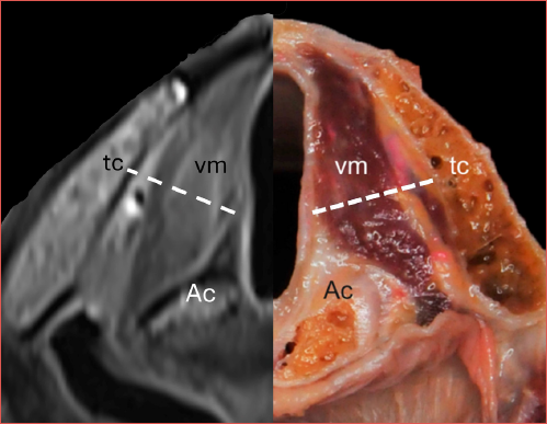
Cover Image
Involvement of the paraglottic space (PGS) in laryngeal cancer has been considered the hallmark of T3 category since the 6th Edition of the TNM Staging System in 2002 (6TNM). Recent evidence has emerged, mainly in the surgical literature, supporting the value of distinguishing the inferior (glottic and subglottic) part of the PGS (iPGS) into anterior and posterior compartments divided by a line tangential to the arytenoid process and perpendicular to the thyroid lamina. The aim of this review was to collect all existing evidence from both the surgical and non-surgical literature supporting the prognostic significance of such a compartmentalisation, in order to propose an amendment to the current 8TNM by distinguishing T3a (glottic tumours with anterior iPGS involvement endoscopically characterised by vocal fold fixation with normal arytenoid movement and radiologically by involvement of the iPGS in front of the above mentioned line) from T3b (glottic tumours endoscopically characterised by impaired arytenoid movement and radiologically by involvement of the iPGS beyond the same line).
Introduction
Paraglottic space (PGS) involvement by glottic tumours has been recognised as a hallmark of advanced disease (radiological cT3 and pT3), together with inner erosion of the thyroid cartilage (again both radiological cT3 and pT3), and arytenoid cartilage fixation (an exclusively endoscopic cT3 criterion taken as a surrogate of major involvement of the crico-arytenoid unit [CAU]) dating back to the Sixth Edition of the AJCC UICC TNM Staging System (6TNM) in 2002 1 and up to the current 8th Edition (8TNM) 2. This is logical if one considers that PGS, in conjunction with the pre-epiglottic space (PES), is a visceral space of the larynx, located deep to the quadrangular membrane, vocal ligament, conus elasticus, and thyro-arytenoid muscle, extending in a vertical direction in between these fibro-muscular structures and the external cartilaginous framework 3. Moreover, its continuity both antero-superiorly with the PES, as well as infero-laterally with the thyro-crico-arytenoid space (TCAS) and extra-laryngeal tissues, make the PGS one of the most probable routes for tumour spread not only within different subsites of the larynx (supraglottis, glottis, and subglottis), but also to the extra-laryngeal anatomical structures of the neck 3.
For all these reasons, the PGS is a crucial pathway for tumour progression in laryngeal cancer, and a thorough evaluation of its status is of paramount importance in staging and treatment selection. This manuscript is aimed at highlighting that there is still ample room to improve our understanding of the real impact of different ways of glottic tumour progression relative to the PGS and describe the nuanced influence of involvement of this visceral space on oncological outcomes after surgical and non-surgical treatments.
Anatomy of the PGS
The PGS is a visceral compartment that is oriented vertically cranio-caudal within the laryngeal framework. It is bounded laterally by the thyroid cartilage, dorsally by the piriform sinus, and medially by the quadrangular membrane, vocal ligament, thyro-arytenoid muscle (TAM), and conus elasticus. This space extends from the level of the superior aspect of the thyroid cartilage to the cricothyroid space. Superiorly and medially it continues without any macroscopic boundary with the PES, forming a unique horseshoe-shaped visceral space that plays a critical role in tumour spread within the supraglottic larynx, from one side to the opposite one.3
Anatomically, the PGS is divided into 2 main portions: the inferior or glottic PGS (iPGS), which lies below the level of the laryngeal ventricle, and the superior or supraglottic PGS (sPGS), located lateral to the quadrangular membrane of the false vocal cord 3. In the iPGS, adipose tissue may create an escape route for tumour spread beyond the larynx. Anteriorly, this may occur in contact with the discontinuous cricothyroid membrane, while posteriorly the tumour may follow a pathway between the lateral cricoarytenoid and cricothyroid muscles toward the CAU 4. To further delineate the posterior region of the iPGS, some authors have proposed the concept of TCAS, which should be herein considered analogous to the posterior part of the iPGS 5. On the top of this, CAU should be clearly defined as the sum of the arytenoid cartilage with underlying crico-arytenoid joint and ipsilateral half of cricoid plate, adjacent lateral and posterior crico-arytenoid muscles, with corresponding blood, lymphatic vessels, and recurrent laryngeal nerve 6. From a pathological perspective, the CAU – along with other cartilage insertion sites of ligaments and muscles, such as the dihedral angle of the thyroid cartilage at the anterior commissure – is subject to ossification due to mechanical stress stimuli. These ossified regions contain adipose and well-vascularised haematopoietic tissue, forming points of minor resistance that favor tumour infiltration. In contrast, hyaline cartilage, with its compact structure and lack of vessels, serves as a natural barrier against neoplastic spread 3,7,8.
Proposal for iPGS compartmentalisation
Recent studies have reported that cT3 glottic lesions extending into the iPGS anterior to a plane passing through the vocal process of the arytenoid and perpendicular to the ipsilateral thyroid lamina have better prognosis compared to those that extend into the posterior compartment, causing CAU involvement with fixation of the corresponding hemilarynx (vocal fold and arytenoid) (Cover figure) 9,10. Given these anatomical relationships, it is evident that tumours invading the posterior iPGS can exhibit cricoarytenoid joint invasion, cricoid plate infiltration, and lateral cricoarytenoid muscle involvement, often leading to arytenoid fixation. These factors contribute to tumour extension beyond the laryngeal framework, promoting spread toward the hypopharynx and extra-laryngeal neck compartment 10. Furthermore, extra-laryngeal involvement of neurovascular structures, such as the recurrent laryngeal nerve, branches of the superior and inferior laryngeal arteries and veins, and associated lymphatic vessels, may facilitate a cranio-caudal tumour spread with lymph node metastases 10,11.
Therefore, the distinct anatomical and histopathological nuances of the anterior vs posterior iPGS and CAU play a pivotal role in tumour progression. Several studies have laid the scientific foundation for the compartmentalisation of the iPGS and established its prognostic significance. A comprehensive understanding of this anatomy not only enhances surgical decision-making and improves the effectiveness of non-surgical laryngeal preservation strategies, but also has the potential for ensuring optimal oncological and functional outcomes in patients with glottic cancer.
Differentiating anterior vs posterior iPGS tumour involvement by endoscopy and imaging
The literature consistently highlights that arytenoid fixation is one of the most reliable predictive factors for posterior iPGS invasion and CAU involvement. Thus, arytenoid fixation, which manifests as immobility of the arytenoid cartilage on endoscopic exam, is often used as a clinical marker to anticipate the extent of tumour spread, particularly to the posterior laryngeal structures 10,12-16.
Various authors, including Katilmis et al. 17 and Succo et al. 18, have reported several causes of arytenoid fixation. These include supraglottic carcinomas extending downward from the sPGS and immobilising the arytenoid due to the weight of the tumour without actual invasion, glottic cancers invading the posterior iPGS and extending toward the CAU, glottic-subglottic carcinomas directly invading the cricoarytenoid joint, glottic carcinomas with direct involvement of intrinsic laryngeal muscles inserting on the arytenoid, and tumours infiltrating the recurrent laryngeal nerve with ensuing laryngeal palsy. Given these various possible mechanisms of arytenoid fixation, it is clear that proper treatment planning is imperative, particularly when considering conservative surgery. It is thus fundamental to assess the underlying cause of arytenoid fixation using a combination of comprehensive endoscopic evaluation and imaging 7,17-19.
A key study by Marchi et al. 20 demonstrated a strong and statistically significant association between arytenoid fixation and radiological evidence of posterior iPGS involvement with a p value of less than 0.0001. Notably, arytenoid fixation acted as a predictor of posterior extension, with a specificity of 99%, sensitivity of 50%, positive predictive value of 93%, and negative predictive value of 84%.
Another important contribution was from Fermi et al. 21 who prospectively compared endoscopic, radiological, and pathological findings in patients with glottic cancer undergoing open partial horizontal laryngectomy (OPHL) types II-III 22 or total laryngectomy (TL). Their findings demonstrated that endoscopic evaluation is reliable for identifying histopathological involvement of the iPGS. Endoscopic evaluation achieved a sensitivity of 89.1%, specificity of 87.5%, positive predictive value of 89.1%, and negative predictive value of 87.5%, underscoring its utility in clinical practice. However, for patients with reduced or fixed arytenoid mobility, the diagnostic values were 78.9%, 88.1%, 65.2%, and 93.7%, respectively. This highlights that endoscopy remains a useful tool for clinical staging of T3 laryngeal cancer.
Lucioni et al. 23 reported that endoscopic findings of a reduced (impaired or absent) vocal cord motility proved to be more sensitive, with better positive and negative predictive values, but less specific than the radiological finding of complete arytenoid sclerosis in detecting infiltration of the arytenoid cartilage. However, a large multi-institutional study published by Ferrari et al. 24 on behalf of the ARYFIX Collaborative Group evaluating 366 videolaryngoscopies and their rating from 22 expert Otorhinolaryngologists, showed concordance of clinical assessment in an astonishingly low 22.7% of cases. Both endoscopic intra- and inter-observer concordance were exceedingly weak, thus prompting a timely scrutiny of the clinical implications of vocal fold/arytenoid impaired mobility/fixation and the need for more objective proxy methods to quantify clinical findings 25,26.
Cross-sectional imaging is the natural complement to clinical evaluation in laryngeal cancer pre-treatment assessment. Imaging, in fact, may significantly contribute to stratifying anterior vs posterior iPGS invasion and detect clinically occult extra-laryngeal extent. While the natural anatomical width of the posterior iPGS generally allows visualisation and thus direct evaluation of its relationships with the tumour, involvement of the anterior iPGS, which is thinner and often less visible, may only be indirectly inferred.
In most cases, computed tomography (CT) is the modality used for glottic tumour staging. However, in spite of optimised acquisition protocols, CT may prove insufficient in discriminating tumour from muscle and oedema, especially if the administration of contrast does not produce significant density contrast between such entities. In these circumstances, magnetic resonance imaging (MRI) proves to be a valuable complement: the multiparametric nature of this technique, resulting in high contrast resolution, increases the possibility to map tumour spread in greater detail; furthermore, the accuracy of MRI may be boosted by the use of surface coils. Ravanelli and coworkers 14,16,27 demonstrated that MRI with surface coils accurately assesses the involvement of the posterior iPGS and CAU (Figs. 1-2). In the presence of clinically detected arytenoid fixation, MRI assessment of cartilage signal intensity is crucial: T2 intermediate signal and restricted diffusion on diffusion weighted imaging indicate invasion, whereas their absence suggests mass effect and/or inflammatory reaction. When the tumour involves the iPGS, contrast resolution of MRI may also be exploited to detect extra-laryngeal spread through the narrow corridor between the thyroid cartilage superiorly, and cricoid cartilage, lateral cricoarytenoid and cricothyroid muscles inferiorly.
Moreover, it should be emphasised that, in a series including 237 CT or MRI scans assessed by 6 radiologists, Ferrari et al. 24 found a low intra-observer variability, but quite weak inter-rater agreement. This observation highlights the importance to include dedicated head and neck radiologists in the multidisciplinary team and to always correlate state-of-the-art imaging with an adequate endoscopic evaluation associated with videorecording.
Evidence supporting iPGS compartmentalisation from a surgical perspective
About 20 years ago, Peretti et al. 28 first compared rates of disease-free survival (DFS), ultimate local control with laser surgery alone (LCL), and laryngeal preservation (LP) between pT2 and pT3 with anterior iPGS invasion treated by carbon dioxide (CO2) transoral laser microsurgery (TOLMS) 29. The survival outcomes of the few patients who were in the cT2/pT3 category (tumours with impaired vocal cord but normal arytenoid mobility due to involvement of the anterior iPGS) showed statistically significant differences compared to the entire cT2/pT2 group (2-year DFS 16.7%, LCL 16.7%, and LP 16.7% for pT3 vs 5-year DFS 80.5%, LCL 84.7%, and LP 93.3% for the whole pT2 group).
In 2010, Vilaseca et al. 9 reported their oncological results in 147 patients treated by CO2 TOLMS for pT3 glottic and supraglottic tumours, stratifying the cohort based upon PES involvement, infiltration of the thyroid cartilage, and arytenoid fixation. The authors reported a 5-year overall survival (OS), disease-specific survival (DSS), and LP of 73.1%, 86.3%, and 51%, respectively for the entire cohort. Moreover, their multivariate analysis highlighted the negative impact of vocal cord fixation (odds ratio [OR] = 2.586; 95% confidence interval [CI] = 1.225-5.457; p = 0.013) as an independent significant predictor of recurrence, organ preservation (OR = 0.184; 95% CI = 0.082-0.411; p = 0.000), and function preservation (OR = 0.198; 95% CI = 0.090-0.435; p = 0.000).
In the same year, Peretti et al. 30 reported a series of 120 glottic tumours, involving 109 pT2 patients (56 with normal, 53 with impaired vocal fold mobility) and 11 pT3 patients with anterior iPGS involvement, all treated by CO2 TOLMS. Five-year DSS, LCL, and LP were 98.3%, 85.6%, and 96.1% for pT2 and 100%, 71.6%, and 72.7% for pT3, thus confirming the worse outcomes for the latter category. In a subsequent study involving 89 T2-T3 glottic tumours treated by TOLMS, the same authors 31 found that anterior iPGS involvement was also a negative prognostic factor for LCL. However, it did not significantly impact the LP rate, as open conservative salvage surgery remained a viable option for this subset of patients.
Vilaseca et al. 32 further confirmed through logistic regression and chi-square automatic interaction detection tree analysis that invasion of the iPGS was independently correlated with local recurrence (hazard ratio [HR] = 2.42, CI 95%= 1.41-4.15; p = 0.001) and LCL (HR = 0.25, CI= 0.14-0.43; p < 0.001). This observation is supported by Ansarin et al. 33 who analysed 590 patients with cTis-T3 glottic cancers treated by TOLMS with curative intent. Specifically, their study confirmed the negative oncologic outcomes of TOLMS for recurrence-free survival (RFS) in pT3 lesions with arytenoid fixation, as previously reported in the literature. Arytenoid cartilage invasion has also been identified as an independent predictor of local recurrence (HR = 6.5; 95% CI = 2.1-26.6) in the cohort reported by Chang et al. 34.
In the study by Piazza et al. 35 which first analysed the different laryngeal isoprognostic zones in the context of TOLMS, the pT3 category with anterior iPGS invasion had a significantly increased risk of local recurrence (HR = 9.2) in comparison to T1-T2 tumours. This subcategory also showed a significantly reduced probability of achieving LCL (HR = 73.6) and LP (HR = 6.4), underscoring the difficulty of managing tumours within the iPGS by TOLMS alone. The most recent update of the isoprognostic zones proposed by the multi-institutional effort of Marchi et al. 36, which assessed a larger cohort of 637 pT2-T3 glottic tumours treated by TOLMS, highlighted that tumours involving the posterior iPGS had significantly poorer DSS (HR = 3.24, 95% CI = 1.01-10.42; p = 0.05) and LCL (HR = 4.03, 95% CI = 1.77-9.18; p < 0.001), as well as a higher probability of requiring TL (HR = 3.70; 95% CI = 1.33-10.32; p = 0.012) compared to other tumour categories. This study concludes that involvement of the posterior iPGS with arytenoid fixation is associated with worse outcomes for all endpoints, underlining that TOLMS alone as a treatment for this particular group of lesions should be considered with great caution. Recent evidence has shown that a selected group of patients with anterior and/or posterior T3 treated by upfront TOLMS may have therapeutic benefit from adjuvant (chemo-)radiation [(C)RT] in the presence of close or R1 margins that are not amenable to wider surgical resections due to anatomical constraints (e.g. tumours reaching but non involving the thyroid or cricoid cartilages) 37.
Vilaseca et al. in 2021 reviewed 262 patients with locally advanced (pT3-T4a) glottic and supraglottic tumours treated by TOLMS 38. In multivariate analysis, anterior (HR = 0.278, 95% CI = 0.128-0.605; p = 0.001) and posterior (HR = 0.269, 95% CI = 0.115-0.630; p = 0.003) iPGS invasion were independent factors of reduced LCL. Furthermore, anterior (HR = 3.613, 95% CI = 1.537-8.495; p = 0.003) and posterior (HR = 5.196, 95% CI = 2.167-12.455; p < 0.001) iPGS involvement were also independent predictors for TL. Patients with posterior iPGS involvement did not differ in DSS from those without posterior iPGS involvement, but did significantly in 5-year laryngectomy-free survival (LFS).
More recently, several studies have focused on iPGS compartmentalisation into an anterior and posterior portion even through an OPHL perspective. It has been thus demonstrated that cT3 lesions extending into the anterior iPGS behave better than those involving the posterior compartment even when treated by open partial laryngectomies 16. In particular, Succo et al. 10 analysed 479 patients with laryngeal cancer, treated by different types of OPHLs, categorising them into 4 groups: I (anterior pT3 with normal arytenoid mobility), II (posterior pT3 with impaired/absent arytenoid mobility), III (anterior pT4a with extra-laryngeal extension and normal mobility), and IV (posterior pT4a with extra-laryngeal extension and impaired/absent mobility). Five-year OS, DSS, DFS, local control (LC), LFS, and laryngo-oesophageal dysfunction-free survival (LEDFS) were significantly better in anterior pT3-T4 tumours (subcategories I and III) compared with the corresponding pT3-T4 posterior ones (subcategories II and IV). Kaplan-Meier estimates of 5-year oncologic outcomes stratified according to the subcategories I and III (anterior tumours) were respectively OS, 95% vs 82.9%; DSS, 97% vs 93.6%; DFS, 91.5% vs 74.6%; LC, 96% vs 78.1%; LFS, 93% vs 77.2%; and LEDFS, 93.1% vs 70.4%. The same 5-year oncologic outcomes stratified according to the subcategories II and IV were respectively OS, 82% vs 79.9%; DSS, 90.7% vs 86.5%; DFS, 81.2% vs 64.2%, LC, 89.1% vs 81.6%; LFS, 77.7% vs 64.1%; and LEDFS, 76.6% vs 64.7%. Moreover, tumours with posterior iPGS involvement were at a higher risk of neck metastases and the rate of pathologic lymph nodes with extranodal extension was also significantly higher in posterior compared to anterior pT3 glottic carcinomas (OR 2.03) 10.
Del Bon et al. 15 analysed 85 patients with pT3-T4a laryngeal cancer treated by OPHL, dividing them into anterior and posterior lesions according to the previously mentioned definition. Five-year OS, DSS, RFS, and LFS in patients with anterior pT3-T4a cancers were 91%, 94.1%, 72.6%, and 70.2%, respectively. Conversely, in case of posterior pT3-T4a lesions, 5-year OS, DSS, RFS, and LFS were 60.3%, 66.3%, 49.1%, and 52%, respectively. Multivariate analysis confirmed that posterior tumour extension was a significant adverse prognostic factor for OS (HR = 3.10, 95% CI = 1.23-7.80; p = 0.02), DSS (HR = 9.10, 95% CI = 2.00-41.43; p = 0.004,), and RFS (HR = 2.51, 95% CI = 1.12-5.64; p = 0.03).
In the study by Lucioni et al. 5, pT3-T4 lesions with involvement of the TCAS (i.e. posterior iPGS) treated by conservative surgery showed a higher recurrence rate (22% vs 8%) and lower DFS (mean 24.1% ± standard deviation [SD] 23.8 vs 31.9% ± SD 28.6, p = 0.06), compared to tumours without TCAS involvement.
De Vincentis et al. 39 in 2022 presented a series of 170 pT2-T4a tumours treated by OPHL type II 22. The 5-year OS rates were 80.9%, 79.3%, and 70.4% for T2, T3, and T4, respectively. DSS rates were 90.4%, 85.3%, and 77.4%. Comparison of outcomes for anterior vs posterior T3-T4a showed a significant reduction in survival outcomes for the latter category. OS, DSS, RFS, and LFS for anterior vs posterior T3 were 86.6%, 89.7%, 92%, and 87% vs 55.5%, 70%, 55.5%, and 55.5%, respectively. OS, DSS, RFS and LFS for anterior vs posterior T4a were 79.1%, 83.3%, 70.8%, and 79.1% vs 42%, 57.8%, 57.1%, and 57.1%, respectively. Uni- and multivariate analysis confirmed that posterior tumour extension was a significant prognosticator for all endpoints: OS (HR = 5.27, 95% CI = 2.64-10.53; p < 0.001), DSS (HR = 4.41, 95% CI = 1.97-9.84; p < 0.001), RFS (HR = 5.04, 95% CI = 2.54-10.01; p < 0.001), and LFS (HR = 5.92, 95% CI = 2.92-11.98; p < 0.001).
In contrast, in the study by Marchi et al. 40, which analysed 149 patients with pT3-T4 laryngeal cancer treated with upfront TL, no significant differences in survival outcomes were found when comparing tumours with anterior vs posterior PGS involvement. In fact, 5-year OS and DSS for anterior vs posterior tumours did not significantly differ in either uni- or multivariable analysis. This observation is not unexpected, as TL radically encompasses the entire PGS thereby neutralising the discrepancies in biological behaviour and pathways of spread of anterior and posterior iPGS cancers that are observed in more conservative surgical approaches such as TOLMS and OPHLs.
Evidence supporting iPGS compartmentalisation from the non-surgical therapeutic modality perspective
There is little or no data about iPGS compartmentalisation or isoprognostic zones and outcomes after RT for glottic cancer. There are several reasons for this lack of data: 1) contrary to surgical series, which can analyse and report pTNM staging, RT series have to rely on non-pathologic, clinical or radiological features that are often subjective; 2) unlike surgical resection, where a precise definition of tumour extent is crucial for the decision of the “technical resectability” and the type of surgical procedure to be performed (e.g. TOLMS vs OPHL vs TL), the possibility to irradiate laryngeal tumours is not affected by the precise tumour extent within the larynx; 3) much of the available data are from the pre-intensity modulated RT era, during which patients were treated with 2D RT without a precise delineation (thus a precise localisation) of the gross tumour volume on planning images.
With this caveat in mind, in glottic tumours treated by 2D RT, iPGS infiltration has been identified as a significant prognostic factor 41-44. Therefore, a more intense treatment with concomitant CRT has been shown to provide better LC compared to RT alone for this subset of T3 tumours 16,45.
Whether posterior iPGS infiltration is worse than anterior involvement after RT is unknown. However, vocal cord impaired mobility (cT2) 46 or fixation (cT3) have been traditionally acknowledged as adverse prognostic factors following RT alone 46,47.
The prognostic implication of vocal cord/arytenoid impaired mobility or fixation in patients treated with RT is often unclear because the aetiology of this clinical sign may be difficult to discern without histopathologic confirmation, whether there is mass effect limiting the movement of the arytenoid cartilage or a true infiltration of the CAU. It is also unclear whether improved LC after RT in former T2a vs T2b, and in overall T2 compared to T3, reflects a progressive increase in the target tumour volume or simply a worse prognosis associated with tumour involving the posterior iPGS. In this context, it has been shown in a randomised controlled trial that hyperfractionated RT (allowing an increase in the radiation dose) was associated with a better LC (although non-significant) for T2 glottic cancers 48. The prognostic impact of tumour volume has also been documented as a potential surrogate of the extent of iPGS infiltration, and perhaps also indirectly of CAU infiltration, in a Canadian study of 319 patients with T3 glottic tumours from 7 centres 49. Almost half of the patients received concomitant CRT. Each 1 cm3 increase in tumour volume was associated with an HR of 1.07 (95% CI, 1.03-1.11) for OS and of 1.04 (95% CI, 1.01-1.07) for DFS.
Altogether, although less precise and accurate than the data from the surgical series, the evidence coming from RT series also suggests that somehow integrating the tumour volume and/or the exact laryngeal tumour localisation within the iPGS could possibly refine staging and prognosis, and thus the selection for non-surgical treatment. Whether this knowledge would help deciding between RT alone or more intense concomitant CRT, however, remains unknown.
Similar to RT, the role of systemic therapy in the context of laryngeal cancer with iPGS involvement is uncertain. Several randomised trials have been conducted in the past, investigating organ preservation in patients with locally advanced laryngeal cancer. Patients were included in the studies based on TNM classification, specifically Stages III and IV, without any specific information about the iPGS involvement. In fact, even though iPGS was included in the TNM classification for the first time in 2002 1 as a hallmark of T3 category, it has been and still continues to be considered as a single entity, without distinguishing between anterior vs posterior compartment or accounting for arytenoid mobility impairment. The efficacy of systemic therapy can be therefore only indirectly inferred by considering arytenoid fixation as a surrogate of posterior iPGS involvement. In organ-preservation trials, systemic therapies were administered either concurrently with RT (platinum-based CRT) or sequentially, as induction chemo- (CHT) or immunotherapy followed by RT, with or without concomitant CHT. In the RTOG 91-11 trial, 3 different treatment approaches were evaluated: sequential therapy, CRT, and RT alone 45. Post-hoc analyses examined local failure rates in patients with T3 disease, stratified by the presence of vocal cord mobility, classified as normal or impaired 50. Among patients with vocal cord fixation, the lowest rate of local failure was observed with CRT (28.5%), compared with sequential therapy (cisplatin + 5-FU [PF] followed by RT) (49.4%) and RT alone (44.1%). In contrast, among patients without vocal cord fixation, local failure rates were lower with either induction PF (29%) or CRT (29%), but remained higher with RT alone (49%). These findings suggest that the efficacy of systemic therapy may be influenced by vocal cord mobility (and therefore by the site of tumour involvement in terms of anterior iPGS with vocal cord fixation but normal arytenoid mobility vs posterior iPGS with vocal cord and arytenoid fixation), highlighting its potential role in treatment planning. A prior randomised trial directly comparing induction PF with TL in patients with T3 laryngeal cancer and vocal cord fixation, further highlighted these differences 51. Both DFS and OS were significantly better with upfront surgery, and the trial was terminated early due to poor accrual, confirming the limited activity of sequential therapy in patients with fixed vocal cords. Recent trials have used return of vocal cord mobility as a marker of response to induction CHT to identify patients who are suitable for organ-preservation approaches 52-54. In the GORTEC 2000-01 trial, patients with Stage III-IV laryngeal and hypopharyngeal cancers were randomised to receive induction CHT with docetaxel, cisplatin, and 5-FU versus PF alone. The triplet regimen achieved a higher vocal cord remobilisation rate (42.7%) compared with the PF regimen (29.1%).
In a retrospective, multi-institutional, collaborative study on 406 patients with T2-T4 laryngeal tumours with impaired mobility (ranging from pure vocal cord impaired mobility to complete hemilaryngeal fixation), non-surgical organ preservation strategies performed better than OPHLs with or without adjuvant (C)RT only in presence of high burden neck disease (N2-N3). Organ preservation surgery in terms of OPHL, on the other hand, performed better than a non-surgical approach in this subset of patients (most of whom had some indirect evidence of posterior iPGS involvement) for all the oncologic outcomes considered (5- and 10-year OS, DSS, RFS, time to distant recurrence, and cumulative incidence of cancer-unrelated deaths) 55.
More recently, immunotherapy, either alone 56 or in combination with CHT 57, has emerged as a promising option for head and neck cancer, both in locally advanced and in relapsed/metastatic disease. In KEYNOTE-689, 22.3% of 363 patients with Stages III-IVA laryngeal cancer received pembrolizumab. A major pathological response was reported in 13.7% of the entire cohort; however, no specific details were provided for laryngeal and hypopharyngeal cancers, nor was a more detailed analysis correlating disease site, stage, and immunotherapy activity reported 56. Preliminary results of the ICoLP trial were presented at the 2023 ASCO meeting by Ferrarotto and coworkers. Patients with Stage II-III disease who were candidates for larynx preservation were treated with pembrolizumab in combination with cisplatin and docetaxel. A total of 24 patients were enrolled, 54.2% of whom had T3 disease. Among the 23 evaluable patients, complete clinical remission was observed in 52%, while 77.3% achieved a pathological complete remission that lasted at least 1 year in 5 patients. In another phase II trial, toripalimab was evaluated in combination with cisplatin and paclitaxel, followed by platinum-based CRT and adjuvant toripalimab for 8 cycles 57. The study enrolled 27 patients with Stage III-IV laryngeal or hypopharyngeal cancer. Notably, 33% of patients had T4a disease and 48.1% had T4b disease. The primary endpoint was achieved, with a 3-month LP rate of 88.9%. The authors emphasised that the activity of CHT-immunotherapy depends more on pre-existing functional impairment of the larynx rather than on tumour extension. Based on the evidence from these 3 recent studies, it is likely that immunotherapy will play a role in the future management of advanced laryngeal and hypopharyngeal cancer, even though, at present, its contribution in treatment of tumours infiltrating the iPGS is still unclear.
Conclusions
Based on a review of the existing evidence from both the surgical and non-surgical literature, it seems reasonable to assume that:
- cT3 glottic tumours with anterior iPGS involvement (endoscopically characterised by vocal fold fixation with normal arytenoid movement and radiologically by involvement of the iPGS anterior to a line tangential to the arytenoid process and perpendicular to the thyroid lamina) have better prognosis and may be defined as cT3a;
- cT3 glottic tumours involving the posterior iPGS and CAU (endoscopically characterised by impaired arytenoid movement and radiologically by involvement of the iPGS posterior to a line tangential to the arytenoid process and perpendicular to the thyroid lamina) have poorer prognosis and may be defined as cT3b.
As a consequence, for pT criteria, we propose that:
- pT3a glottic tumours are those with full-thickness TAM invasion, infiltration of the iPGS anteriorly to a plane passing through the arytenoid vocal process, or inner thyroid cartilage plate infiltration;
- pT3b glottic tumours are those with full thickness TAM and iPGS invasion posteriorly to a plane passing through the arytenoid vocal process.
Conflict of interest statement
The authors declare no conflict of interest.
Funding
This research did not receive any specific grant from funding agencies in the public, commercial, or not-for-profit sectors.
Author contributions
All authors contributed to the interpretation of the data, critically revised the manuscript for important intellectual content, and approved the final version of the manuscript.
Ethical consideration
Not applicable.
History
Received: October 15, 2025
Accepted: March 9, 2026
Figures and tables
Figure 1. Axial T2-weighted (A) and unenhanced T1-weighted (B) images of squamous cell carcinoma of the right vocal cord (T) invading the most anterior part of the iPGS (effacement of fat signal, thin white arrow). In Figure 1B, a focal low signal in the anterior right thyroid lamina can be seen (thick white arrow), corresponding to high signal in Figure 1A (finding consistent with reactive chondritis). The lesion lies in front of the dashed line dividing the anterior and posterior compartments of the iPGS. Black arrowheads, posterior paraglottic fat. A: arytenoids; vm: vocal muscle.
Figure 2. Axial T2-weighted MRI of a right glottic tumour (t) with endoscopically fixed vocal cord and arytenoid, involving both the anterior and posterior iPGS through the interrupted white line tangential to the arytenoid (a) vocal process and perpendicular to the thyroid cartilage lamina (ThC). On the left healthy side, the normal fat tissue filling the iPGS lateral to the thyroarytenoid vocal muscle (vm) is shown for clarity.
References
- Greene F, Page D, Fleming I. AJCC Cancer Staging Manual. 6th ed. New York: Springer; 2002.
- Amin M, Edge S, Greene F. AJCC Cancer Staging Manual. 8th ed. New York: Springer; 2017.
- Lucioni M. Practical guide to neck dissection: focusing on the larynx. Heidelberg, Germany: Springer; 2013.
- Reidenbach M. The paraglottic space and transglottic cancer: anatomical considerations. Clin Anat. 1996;9:244-251. doi:https://doi.org/10.1002/(SICI)1098-2353(199699:4<244::AID-CA5>3.0.CO;2-E
- Lucioni M, Lionello M, Guida F. The thyro-cricoarytenoid space (TCAS): clinical and prognostic implications in laryngeal cancer. Acta Otorhinolaryngol Ital. 2020;40:106-112. doi:https://doi.org/10.14639/0392-100X-N0373
- Weinstein G, Laccourreye O, Brasnu D. Organ preservation surgery for laryngeal cancer. San Diego, California: Singular Publishing Group; 2000.
- Kirchner J, Som M. Clinical significance of fixed vocal cord. Laryngoscope. 1971;81:1029-1044. doi:https://doi.org/10.1288/00005537-197107000-00005
- Kirchner J, Som M. Clinical and histological observations on supraglottic cancer. Ann Otol Rhinol Laryngol. 1971;80:638-645. doi:https://doi.org/10.1177/000348947108000503
- Vilaseca I, Bernal-Sprekelsen M, Blanch J. Transoral laser microsurgery for T3 laryngeal tumors: prognostic factors. Head Neck. 2010;32:929-938. doi:https://doi.org/10.1002/hed.21288
- Succo G, Crosetti E, Bertolin A. Treatment for T3 to T4a laryngeal cancer by open partial horizontal laryngectomies: prognostic impact of different pathological tumor subcategories. Head Neck. 2018;40:1897-1908. doi:https://doi.org/10.1002/hed.25176
- Iimura A, Itoh M, Terayama H. Anatomical study of meandering and functions of human intralaryngeal artery. Okajimas Folia Anat Jpn. 2004;81:85-92. doi:https://doi.org/10.2535/ofaj.81.85
- Vilaseca I, Bernal-Sprekelsen M. Transoral laser microsurgery for locally advanced laryngeal cancer. Acta Otorrinolaringol Esp. 2012;64:140-149. doi:https://doi.org/10.1016/j.otorri.2012.01.013
- Peretti G, Piazza C, Mora F. Reasonable limits for transoral laser microsurgery in laryngeal cancer. Curr Opin Otolaryngol Head Neck Surg. 2016;24:135-139. doi:https://doi.org/10.1097/MOO.0000000000000240
- Ravanelli M, Paderno A, Del Bon F. Prediction of posterior paraglottic space and cricoarytenoid unit involvement in endoscopically T3 glottic cancer with arytenoid fixation by magnetic resonance with surface coils. Cancers (Basel). 2019;11. doi:https://doi.org/10.3390/cancers11010067
- Del Bon F, Piazza C, Lancini D. Open partial horizontal laryngectomies for T3-T4 laryngeal cancer: prognostic impact of anterior vs posterior laryngeal compartmentalization. Cancers (Basel). 2019;11. doi:https://doi.org/10.3390/cancers11030289
- Ferrari M, Mularoni F, Smussi D. International consensus on laryngeal preservation strategies in laryngeal and hypopharyngeal cancer. Lancet Oncol. 2025;26:E264-E281. doi:https://doi.org/10.1016/S1470-2045(25)00020-8
- Katilmiş H, Oztürkcan S, Ozdemir I. A clinico-pathological study of laryngeal and hypopharyngeal carcinoma: correlation of cord-arytenoid mobility with histopathologic involvement. Otolaryngol Head Neck Surg. 2007;136:291-295. doi:https://doi.org/10.1016/j.otohns.2006.08.022
- Succo G, Cirillo S, Bertotto I. Arytenoid fixation in laryngeal cancer: radiological pictures and clinical correlations with respect to conservative treatments. Cancers (Basel). 2019;11. doi:https://doi.org/10.3390/cancers11030360
- Prades J, Dumollard J, Timoshenko A. Descriptive anatomy of the cricoarytenoid articulation: application to articular dynamics in carcinology. Surg Radiol Anat. 2000;22:277-282. doi:https://doi.org/10.1007/s00276-000-0277-y
- Marchi F, Filauro M, Missale F. A multidisciplinary team guided approach to the management of cT3 laryngeal cancer: a retrospective analysis of 104 cases. Cancers (Basel). 2019;11. doi:https://doi.org/10.3390/cancers11050717
- Fermi M, Lo Manto A, Di Massa G. Paraglottic space invasion in glottic laryngeal cancer: a clinical-pathological study. Laryngoscope. 2023;133:1184-1190. doi:https://doi.org/10.1002/lary.30335
- Succo G, Peretti G, Piazza C. Open partial horizontal laryngectomies: a proposal for classification by the working committee on nomenclature of the European Laryngological Society. Eur Arch Otorhinolaryngol. 2014;271:2489-2496. doi:https://doi.org/10.1007/s00405-014-3024-4
- Lucioni M, Lionello M, Machin P. Sclerosis of the arytenoid cartilage and glottic carcinoma: a clinical-pathological study. Head Neck. 2018;41:72-78. doi:https://doi.org/10.1002/hed.25372
- Ferrari M, Mularoni F, Taboni S. How reliable is assessment of true vocal cord-arytenoid unit mobility in patients affected by laryngeal cancer? A multi-institutional study on 366 patients from the ARYFIX collaborative group. Oral Oncol. 2024;152. doi:https://doi.org/10.1016/j.oraloncology.2024.106744
- Villani F, Paderno A, Fiorentino M. Classifying vocal folds fixation from endoscopic videos with machine learning. Annu Int Conf IEEE Eng Med Biol Soc. 2023;202:1-4. doi:https://doi.org/10.1109/EMBC40787.2023.100340017
- Villani F, Fiorentino M, Federici L. A deep-learning approach for vocal fold pose estimation in videoendoscopy. J Imaging Inform Med. 2026;39:842-852. doi:https://doi.org/10.1007/s100278-025-01431-8
- Maroldi R, Ravanelli M, Farina D. Magnetic resonance for laryngeal cancer. Curr Opin Otolaryngol Head Neck Surg. 2014;22:131-139. doi:https://doi.org/10.1097/MOO.0000000000000036
- Peretti G, Piazza C, Mensi M. Endoscopic treatment of cT2 glottic carcinoma: prognostic impact of different pT subcategories. Ann Otol Rhinol Laryngol. 2005;114:579-586. doi:https://doi.org/10.1177/000348940511400801
- Remacle M, Arens C, Eldin M. Laser-assisted surgery of the upper aero-digestive tract: a clarification of nomenclature. A consensus statement of the European Laryngological Society. Eur Arch Otorhinolaryngol. 2017;274:3723-3727. doi:https://doi.org/10.1007/s00405-017-4708-3
- Peretti G, Piazza C, Cocco D. Transoral CO2 laser treatment for Tis-T3 glottic cancer: the University of Brescia experience on 595 patients. Head Neck. 2010;32:977-983. doi:https://doi.org/10.1002/hed.21278
- Peretti G, Piazza C, Del Bon F. Function preservation using transoral laser surgery for T2–T3 glottic cancer: oncologic, vocal, and swallowing outcomes. Eur Arch Otorhinolaryngol. 2013;270:2275-2281. doi:https://doi.org/10.1007/s00405-013-2461-9
- Vilaseca I, Nogués-Sabaté A, Avilés-Jurado F. Factors of local recurrence and organ preservation with transoral laser microsurgery in laryngeal carcinomas; CHAID decision-tree analysis. Head Neck. 2019;41:756-764. doi:https://doi.org/10.1002/hed.25422
- Ansarin M, Cattaneo A, De Benedetto L. Retrospective analysis of factors influencing oncologic outcome in 590 patients with early-intermediate glottic cancer treated by transoral laser microsurgery. Head Neck. 2017;39:71-81. doi:https://doi.org/10.1002/hed.24534
- Chang C-F, Chu P-Y. Predictors of local recurrence of glottic cancer in patients after transoral laser microsurgery. J Chin Med Assoc. 2017;80:452-457. doi:https://doi.org/10.1016/j.jcma.2017.04.002
- Piazza C, Filauro M, Paderno A. Three-dimensional map of isoprognostic zones in glottic cancer treated by transoral laser microsurgery as a unimodal treatment strategy. Front Oncol. 2018;8. doi:https://doi.org/10.3389/fonc.2018.00175
- Marchi F, Del Bon F, Chu F. Refining prognostic subcategories in intermediate-advanced glottic cancer: a multicentric study on 637 patients treated by transoral laser microsurgery. Oral Oncol. 2025;164. doi:https://doi.org/10.1016/j.oraloncology.2025.107264
- Pinacoli A, Ruiz-Sevilla L, Sampieri C. Optimizing oncologic outcomes in pT3 glottic cancers treated by transoral laser microsurgery: the impact of adjuvant treatment. Acta Otorhinolaryngol Ital. 2025;45:388-398. doi:https://doi.org/10.14639/0392-100X-A1372
- Vilaseca I, Aviles-Jurado F, Valduvieco I. Transoral laser microsurgery in locally advanced laryngeal cancer: prognostic impact of anterior versus posterior compartments. Head Neck. 2021;43:3832-3842. doi:https://doi.org/10.1002/hed.26878
- De Vincentiis M, Greco A, Campo F. Open partial horizontal laryngectomy for T2-T3-T4a laryngeal cancer: oncological outcomes and prognostic factors of two Italian hospitals. Eur Arch Otorhinolaryngol. 2022;279:2997-3004. doi:https://doi.org/10.1007/s00405-021-07238-x
- Marchi F, Missale F, Sampieri C. Laryngeal compartmentalization does not affect the prognosis of T3-T4 laryngeal cancer treated by upfront total laryngectomy. Cancers (Basel). 2020;12:1-18. doi:https://doi.org/10.3390/cancers12082241
- Pameijer F, Mancuso A, Mendenhall W. Can pretreatment computed tomography predict local control in T3 squamous cell carcinoma of the glottic larynx treated with definitive radiotherapy?. Int J Radiat Oncol Biol Phys. 1997;37:1011-1021. doi:https://doi.org/10.1016/s0360-3016(96)00626-8
- Hermans R, Van den Bogaert W, Rijnders A. Predicting the local outcome of glottic squamous cell carcinoma after definitive radiation therapy: value of computed tomography-determined tumour parameters. Radiother Oncol. 1999;50:39-46. doi:https://doi.org/10.1016/s0167-8140(98)00114-5
- Dagan R, Morris C, Bennett J. Prognostic significance of paraglottic space invasion in T2N0 glottic carcinoma. Am J Clin Oncol. 2007;30:186-190. doi:https://doi.org/10.1097/01.coc.0000251403.54180.df
- McCoul E, Har-El G. Meta-analysis of impaired vocal cord mobility as a prognostic factor in T2 glottic carcinoma. Arch Otolaryngol Head Neck Surg. 2009;135:479-486. doi:https://doi.org/10.1001/archoto.2009.47
- Forastiere A, Goepfert H, Maor M. Concurrent chemotherapy and radiotherapy for organ preservation in advanced laryngeal cancer. N Engl J Med. 2003;349:2091-2098. doi:https://doi.org/10.1056/NEJMoa031317
- Wittekind C, Henson D, Hutter R. TNM Supplement. A Commentary on Uniform Use. Springer; 1983.
- Mendenhall W, Amdur R, Morris C. T1-T2N0 squamous cell carcinoma of the glottic larynx treated with radiation therapy. J Clin Oncol. 2001;19:4029-4036. doi:https://doi.org/10.1200/JCO.2001.19.20.4029
- Trotti A, Zhang Q, Bentzen S. Randomized trial of hyperfractionation versus conventional fractionation in T2 squamous cell carcinoma of the vocal cord (RTOG 9512). Int J Radiat Oncol Biol Phys. 2014;89:958-963. doi:https://doi.org/10.1016/j.ijrobp.2014.04.041
- Malik N, Fu R, Hainc N. Association of primary tumor volume with survival in patients with T3 glottic cancer treated with radiotherapy: a study of the Canadian Head & Neck Collaborative Research Initiative. JAMA Otolaryngol Head Neck Surg. 2023;149:103-109. doi:https://doi.org/10.1001/jamaoto.2022.3996
- Olsen K. Reexamining the treatment of advanced laryngeal cancer. Head Neck. 2010;32:1-7. doi:https://doi.org/10.1002/hed.21294
- Richard J, Sancho-Garnier H, Pessey J. Randomized trial of induction chemotherapy in larynx carcinoma. Oral Oncol. 1998;34:224-228. doi:https://doi.org/10.1016/s1368-8375(97)00090-0
- Lefebvre J, Rolland F, Tesselaar M. Phase 3 randomized trial on larynx preservation comparing sequential vs alternating chemotherapy and radiotherapy. J Nat Cancer Inst. 2009;101:142-152. doi:https://doi.org/10.1093/jnci/djn460
- Lefebvre J, Pointreau Y, Rolland F. Induction chemotherapy followed by either chemoradiotherapy or bioradiotherapy for larynx preservation: the TREMPLIN randomized phase II study. J Clin Oncol. 2013;31:853-859. doi:https://doi.org/10.1200/JCO.2012.42.3988
- Pointreau Y, Garaud P, Chapet S. Randomized trial of induction chemotherapy with cisplatin and 5-fluorouracil with or without docetaxel for larynx preservation. J Nat Cancer Institute. 2009;101:498-506. doi:https://doi.org/10.1093/jnci/djp007
- Mularoni F, Marchi F, Gaudioso P. Selected laryngeal squamous cell carcinomas with laryngeal mobility impairment are suitable for curative larynx-preservation treatment. A multi-institutional study on 406 patients from the ARYFIX collaborative group. Oral Oncol. 2025;167. doi:https://doi.org/10.1016/j.oraloncology.2025.107466
- Uppaluri R, Haddad R, Tao Y. Neoadjuvant and adjuvant pembrolizumab in locally advanced head and neck cancer. N Engl J Med. 2025;393:37-50. doi:https://doi.org/10.1056/NEJMoa2415434
- Xiaomin O, Ruiping Z, Wenjun W. Induction toripalimab and chemotherapy for organ preservation in locally advanced laryngeal and hypopharyngeal cancer: a single-arm phase II clinical trial. Clin Cancer Res. 2024;30:344-355. doi:https://doi.org/10.1158/1078-0432.CCR-23-2398
Downloads
License

This work is licensed under a Creative Commons Attribution-NonCommercial-NoDerivatives 4.0 International License.
Copyright
Copyright (c) 2026 Società Italiana di Otorinolaringoiatria e chirurgia cervico facciale
How to Cite
- Abstract viewed - 274 times
- PDF downloaded - 69 times