


Reviews
Vol. 46: Issue 4 (Suppl. 1) - August 2026
Glottic tumours involving the anterior commissure in a horizontal vs vertical mode have different prognosis. Opportunity for refinement of the TNM 8th Edition
Summary
Cover Image
The anterior commissure (AC) represents a unique subsite of the glottic plane due to its peculiar embryological development and 3-dimensional anatomical microstructure. More than the simple junction between 2 vocal cords, in fact, the AC should be viewed as the confluence between the petiolar region and pre-epiglottic space superiorly, the anterior thirds of the false and true vocal cords and related paraglottic spaces laterally, and the conoid ligament inferiorly. These anatomical features have profound impact on the behaviour of laryngeal cancer at the level of the AC. In particular, the neoplastic patterns of growth (on the horizontal plane from one vocal fold to the contralateral one remaining on a purely glottic plane vs a vertical trans-AC extension from the supra- to the subglottis or vice versa) have distinct oncologic implications when treating these lesions by both surgical and non-surgical strategies. Therefore, the aim of this manuscript is to collect all the existing evidence in favour of the necessity to review the 8th Edition of the TNM Staging System by distinguishing tumours that involve the glottic plane without AC extension (cT1a of one and cT1b of 2 vocal cords without AC involvement) from those that encompass the AC. Based on available evidence, the prognostic implication of AC involvement should be considered in upstaging these tumours to T2, subclassifying them into T2a (horizontal involvement of the AC) and T2b (vertical trans-AC extension above and/or below the AC).
Introduction
The anterior commissure (AC) should not be considered as a simple subsite of the glottis but rather as a complex 3-dimensional space, sharing part of its characteristics with the vocal cords, but also having special features that make it a unique site within the larynx. In fact, it is entirely filled with dense homogeneous connective tissue with a low density vascular and lymphatic network, in close relationship with the adjacent visceral compartments, i.e. the pre-epiglottic space (PES) superiorly, the superior (sPGS) and inferior paraglottic spaces (iPGS) laterally, and the loose vessel- and minor salivary and mucous gland-rich subglottic tissue inferiorly. It encompasses different anatomical structures such as the anterior maculae flavae of the 2 vocal ligaments and the thyroid cartilage insertions of the thyroarytenoid muscles (TAM) at the glottic level, the thyro-epiglottic ligament and the anterior portions of the quadrangular membranes at the supraglottic level, the conoid ligament in the subglottis, with part of the inner thyroid perichondrium and thyroid laminae anteriorly (Fig. 1) 1-3.
To date, for cancer staging purposes, the AC has been always considered an integral part of the glottic plane and, therefore, its neoplastic involvement in both the horizontal as well as in the vertical directions has been defined in a rather ambiguous way. For example, while T1a specifically indicates a glottic tumour restricted to one vocal cord without extension to the AC, T1b can be represented by any one of 4 different scenarios: 1) 2 discrete lesions affecting the right and left vocal folds without AC involvement; 2) a tumour originating within the AC without significant extension to the vocal cords; 3) a tumour of one vocal cord extending to the AC; or 4) a horseshoe tumour involving the AC and both vocal cords. The distinct potential prognostic implications associated with these 4 different lesions can cause inhomogeneity in definition and recording of oncologic outcomes. More importantly, a tumour growing cranially and/or caudally from the AC to the supra- and/or subglottis is designated as T2 similar to a laterally located T2 extending superiorly from the middle third of the true vocal cord to the superficial part of the false vocal cord and/or inferiorly to the subglottis. Indeed, each of these lesions present very different biologic behaviours and oncologic outcomes when treated by either surgical or non-surgical modalities.
Therefore, several reasons support modification of the current 8th Edition of the TNM Staging System (8TNM) 4 for tumours involving the AC: 1) these lesions do not seem to be inherently more aggressive from a biological point of view, but are more prone to progression due to specific anatomical features of the AC itself; 2) AC tumours require more aggressive treatments compared to lesions not involving this structure in order to achieve comparable outcomes; 3) incorporating AC involvement into T staging could help resolve the above mentioned ambiguous classification of primary tumours involving this anatomical structure.
The aim of this manuscript is to examine the rationale for a revision of the 8TNM of the larynx, specifically addressing the AC as a distinct anatomic region with unique features derived from its embryological development, and its horizontal vs vertical mode of neoplastic invasion which have important prognostic implications that should be acknowledged by a modern staging system.
Anatomy and embryology of the anterior commissure
The embryological development of the AC is important as a basis for understanding the prognostic implications of its neoplastic involvement. The AC becomes identifiable as a discrete structure around the eighth week of gestation, when the 2 hemilarynges fuse to form a 3-dimensional structure known as the intermediate lamina, or developmental AC, which is rich in elastic fibres and contains blood vessels, the median process, and connective tissue of the conoid ligament. The median process evolves into Broyles’ ligament, which connects deeply with the intermediate lamina and functions as a perichondrium for its dorsal surface, located just above the glottic plane. This ligament is integral to the commissural region, providing structural support 2,5. By the eleventh week of gestation, TAM fibres connect with the median process, reinforced by Broyles’ ligament, integrating them into the commissural region 2,3. By the end of development, the so-called “AC ligament tendon” is composed of a dense network of fibres interwoven in both horizontal and vertical directions, originating from the vocal ligament, conus elasticus, and quadrangular membrane. The tendon fibres insert directly into the hyaline cartilage, with no perichondrium at the insertion site, and chondroid cells are embedded among the collagen fibres. The characteristics acquired by the commissure at the end of development define it as a distinct 3-dimensional region, extending approximately 8.5 mm above and below the glottic plane (Fig. 1). This differentiates it from the classical topographic definition of the glottis, which is delimited by 2 axial planes parallel to the free edge of the true vocal folds, positioned approximately 3 mm above and below the glottic plane (Fig. 2).
Unlike all other laryngeal subsites, where muscle tissue, fat of the PGS, and/or perichondrium are situated between the mucosa and the cartilage, the embryological development of AC results in the mucosa being closer to the cartilaginous framework in this region than in any other part of the larynx, with a separation of only 2-3 mm. This close association, combined with mechanical stress at this site, often leads to focal ossification of the thyroid cartilage, which further compromises resistance to neoplastic invasion. Ossification typically begins in the early fourth decade of life at the level of the AC median process and progresses laterally with aging. At this level, ossification occurs through a Haversian system, which is characterised by the formation of Volkmann’s canals (horizontal, running from inside outward) and Haversian canals (vertically oriented in a cranio-caudal direction). These canals are traversed by blood and lymphatic vessels, forming an anastomotic lympho-vascular network – which is absent in cartilage – that connects the endolaryngeal lumen at the level of the pericommissural region with the prelaryngeal tissues. Therefore, the implication of cartilage ossification is that it facilitates tumour invasion, increasing the risk of tumour spread beyond this natural barrier 6,7.
Evaluation of the anterior commissure and its neoplastic involvement by endoscopy and imaging
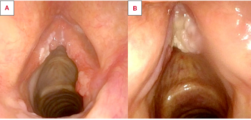
Endoscopic evaluation of laryngeal cancer plays a pivotal role before any type of surgical or non-surgical treatment, and this is especially true in the AC where the precise bidimensional extent of the lesion may greatly impact the choice of therapeutic approach. The need for a major AC resection, for example, may direct treatment of a given tumour towards a non-surgical approach in patients with a high priority for optimal vocal outcomes. In this sense, office-based videoendoscopy under local anaesthesia is rarely precise enough to clearly map the local extent of AC tumours. Rigid endoscopy using angled telescopes under general anaesthesia is therefore considered the state-of-the-art clinical assessment tool for staging these tumours 8. Even though unfavourable laryngeal exposure may negatively impact AC visualisation 9-12, every technical effort should be made to distinguish at least 3 different scenarios: 1) tumours growing from one or both vocal cords towards the AC, without reaching it; 2) tumours growing from one vocal cord across to the contralateral one, involving the AC in a purely horizontal fashion (Cover figure A); 3) tumours passing through the AC in a vertical direction, involving the petiole of the epiglottis, the anterior portions of the false and true vocal cords, and/or the crico-thyroid membrane inferiorly (Cover figure B). Narrow band imaging and other bioendoscopic in vivo imaging tools may be of help in fine-tuning superficial detection of neoplastic spread along one or more of these pathways 13-17.
AC assessment on computed tomography (CT) and magnetic resonance imaging (MRI) is heavily dependent on a correct and optimised scanning protocol and on accurate post-processing. Most studies consider CT as the reference modality 18-27: CT protocols are in fact relatively standardised, typically involving image acquisition approximately 80 seconds after intravenous administration of iodinated contrast medium, either during quiet breathing or with dynamic manoeuvres such as deep inspiration or phonation. CT generates volumetric datasets that must be properly reconstructed. For AC assessment, axial reconstructions parallel to the glottic plane and sagittal reconstructions to evaluate the vertical extension of tumours infiltrating the AC are particularly important 25. Some studies have proposed dual-energy CT to enhance contrast resolution, especially for the detection of cartilage infiltration 28,29 and compared it with MRI with promising results 30.
Conversely, MRI protocols reported in the literature are highly heterogeneous and often not described in sufficient detail 31. Both 1.5 and 3 Tesla systems are employed, and a pilot study has investigated the use of a 7 Tesla scanner 32. Van Egmond, in a systematic review, emphasised that currently available MRI protocols are suboptimal for the staging of early laryngeal cancer, without, however, addressing specific technical details 33. Both neck and surface coils can be used: the latter allow acquisition of images with high spatial resolution and excellent signal-to-noise ratio and are therefore extremely useful for AC assessment 34. High matrix and motion-correction sequences should be used 31,34. The most useful acquisition planes are axial and sagittal. Diffusion-weighted sequences improve the ability to differentiate tumour from inflammatory changes; however, the spatial resolution is limited, and direct acquisition in the sagittal plane is problematic 34.
According to the literature, the normal radiologic thickness of the AC is approximately 1 mm 27, although some variability is accepted (± 0.56 mm). Only one study has described the vertical position of the AC, measuring its distance from the inferior margin of the thyroid cartilage (approximately 11 mm), a parameter relevant for assessing tumour extension toward the cricothyroid space 23.
Most studies address AC neoplastic invasion in a non-specific manner, without further subclassification and with limited description of imaging criteria. The study by Benazzo et al. 22 represents the only attempt to radiologically stage AC infiltration, proposing a detailed mapping system that distinguishes presence of tumour, superficial involvement, thyroid cartilage invasion, and extra-laryngeal spread. The reported sensitivity and specificity were, respectively, 73% and 77% for tumour detection, 25% and 96% for superficial involvement, 59% and 82% for thyroid cartilage invasion, and 100% and 88% for extra-laryngeal spread. This study highlighted the considerable underestimation of AC involvement by CT, and the authors suggested that MRI may be useful in equivocal cases. Notably, vertical tumour extension was not included in their proposed mapping system; in addition, no cut-off thickness value was reported for the superficial involvement of the AC. According to Cristalli et al. 26, the cut-off thickness value on axial scans to discriminate the presence of cancer at histological examination of the AC is 3.6 mm (positive and negative predictive values were 93% and 87%, respectively).
Hartl et al. 20 found that CT had the lowest sensitivity for cartilage invasion (10%); this result must be however interpreted with caution, as the study included 236 patients, of whom only 8% had inner cortex thyroid cartilage involvement. Nonetheless, it is generally accepted that CT is not reliable for staging T3 tumours with inner thyroid cartilage infiltration: in fact, the most specific radiological sign for thyroid involvement is complete replacement of the cartilage by tissue exhibiting the same imaging characteristics as the tumour, whereas the least specific sign is cartilage sclerosis.
Barbosa et al. 19 reported that CT had an overall accuracy of 75%, and when stratified by T category was 62% for T2, 74% for T3, and 88% for T4 lesions. However, they did not specify whether extra-laryngeal extension occurred through the cartilage or via the cricothyroid space.
Taha et al. 35 found a good performance of diffusion-weighted MRI sequences in detecting infiltration of the inner cortex (sensitivity 93%, specificity 88%) and outer cortex (sensitivity 85%, specificity 85%) of the thyroid cartilage. Additional studies highlighting the superiority of MRI over CT in this context are those from Beitler et al. 36, Li et al. 37, and Adolphs et al. 38.
Some authors have investigated the potential of ultrasound (US) for staging laryngeal lesions since the AC may represent an appropriate target for this technique owing to its superficial location 39,40. However, US is highly operator-dependent and far from being a routine standard.
New proposal for staging of glottic tumours involving the anterior commissure
Our proposal for updating the cT classification of glottic tumours involving the AC is based on the distinct embryologic and anatomic issues that influence clinical behaviour and prognosis of these tumours, and include the following:
- cT1a of one vocal cord behaves in a similar way as cT1b of 2 vocal cords without AC involvement. The latter should be, however, maintained as cT1b since, from the probabilistic point of view and considering the field of cancerisation phenomenon, these lesions have a slightly higher tendency to recur compared to unilateral cT1a;
- cT1b of both vocal cords with horizontal involvement of the AC purely in the glottic plane behaves worse than cT1a and cT1b without AC involvement and should be therefore upstaged to cT2a (Cover figure A, Fig. 3);
- cT2 of the AC extending vertically above and below the glottic plane (trans-AC tumours) (Cover figure B, Figs. 4A and B) behaves far worse than actual cT1b and superficial lateral cT2 of the mid-cord not involving the TAM and with normal vocal fold mobility. Additionally, in trans-AC tumours, minimal involvement of the thyro-epiglottic ligament and petiole of the epiglottis, PES and/or inner portion of the thyroid cartilage and loose subglottic tissue cannot be easily ruled out even by state-of-the-art imaging, with the need for targeted (and invasive) biopsies under general anaesthesia. These tumours should therefore be upstaged, according to our proposal, at least to the cT2b category and specifically need extra scrutiny in the diagnostic phase. In fact, if involvement of PES and/or inner thyroid cartilage can be clearly demonstrated, these elements will upstage such tumours to cT3, while through and through extension to the extra-laryngeal tissues will be designated cT4a, as per current criteria (Fig. 5).
Evidence supporting the new proposal for anterior commissure staging from the surgical and non-surgical perspectives
The above mentioned anatomical and embryological principles explain why the inner cortex of the thyroid cartilage at the AC level may be microscopically infiltrated more often than expected 41. Consequently, an early-stage carcinoma involving the AC has the potential to penetrate the cartilage, with significant implications in upstaging a T1-T2 lesion to a T3 or even a T4a tumour 42 (Fig. 5). Therefore, patients affected by cT2-T3 glottic tumours involving the AC are, among all laryngeal cancers, those most frequently understaged (in approximately 16-18% of cases) 43. Following surgical resection, this understaging is clearly associated with a higher risk of positive surgical margins and a consequent decrease of disease-free and disease-specific survivals 44,45. Moreover, the complex anatomical structure of the AC is likely to significantly impact on tumour spread in a number of ways that should be taken into account by a modern TNM staging system of laryngeal cancer. In fact, involvement of the AC by a glottic tumour has the potential to open up pathways of spread in at least 3 major directions: horizontally (from one vocal cord to the other, along the glottic plane), vertically (from cranial to caudal or vice versa, to the supra- and/or subglottis), and postero-anteriorly (from inside to outside, involving the cartilaginous framework of the thyroid cartilage, or at the thyro-hyoid and/or crico-thyroid membrane levels cranially and caudally).
There is general agreement, even in the early glottic cancer scenario, that AC involvement can negatively impact both oncological and functional outcomes, regardless of the therapeutic strategy adopted. Some authors have recently reported AC involvement as a factor that significantly influences the risk of local recurrence and/or the organ preservation rate (Tables I and II) 46-67. Conversely, a limited number of other authors 68-75 have not found an independent role of AC involvement on local relapse. This variability can be explained by a number of factors like variation in the clinical definition of AC, different levels of detail in clinical, endoscopic, and radiologic evaluation in the pre-treatment setting, distinctive features and limitations related to each single therapeutic modality, different biological tumour behaviours in distinct histopathologic entities, as well as variations in the rigor of the follow-up policy.
A small number of systematic reviews and meta-analyses 76-78 have attempted to provide more concrete data on the prognostic role of AC involvement in early glottic cancer. Unfortunately, unacceptable rates (> 50%) of heterogeneity among selected studies limit the validity of results and feasibility of pooled analyses 65. Moreover, since AC involvement may be correlated to other clinical variables (i.e. T category), its independent prognostic role can only be assessed within studies that control for other confounders, and this is usually not the case within published meta-analyses 76-78.
Upon closer review of the available literature, the differences in terms of recurrence rates in tumours with horizontal vs vertical trans-AC extension have mostly been observed in studies that precisely address these potential different patterns of AC involvement. These studies have shown that, in particular, the cranio-caudal extension of the commissural tumours towards the infrapetiolar area and/or the crico-thyroid membrane is a significant variable affecting local control and organ preservation (Tab. III) 46,48,50,52,53,55,79,80. This has recently prompted recommendations for a more aggressive management of these tumours 53,55,61 and a special type of endoscopic cordectomy (Type VI) has been also codified to appropriately modify transoral resection of AC tumours with vertical extension 81, in addition to the 5 previously described within the European Laryngological Society classification 82. Notably, Type VI cordectomy is the most aggressive transoral procedure conceived for tumours located at the glottic level and encompasses the anatomical boundaries previously described as 3-dimensional limits of the AC (from the petiole of the epiglottis above, to the crico-thyroid membrane below, including the anterior thirds of both false and true vocal cords).
Wu et al. 83 assessed 31 glottic carcinomas with AC involvement in patients who underwent partial or total laryngectomy (TL). The specimens were studied by axial serial sections parallel to the vocal cords and analysed to explore the characteristics and potential invasion pathways of glottic carcinomas with AC involvement. In this study, the involvement of the intermediate lamina of the thyroid cartilage was significantly associated with invasion of the vocal muscle in the AC region (p < 0.05). Additionally, Rifai et al. 84 performed a histopathological examination of the AC using whole organ sections in 30 randomly selected specimens affected by AC tumours. They found that microscopic involvement of the thyroid cartilage without penetration was detected in all sections analysed. These findings indicate that tumours in this region have a higher risk of presenting microscopic foci of infiltration in the AC, which may be undetectable prior to surgery and ensuing histopathological examination.
Therefore, tumours originating from the AC, in contrast to those from other laryngeal subsites, necessitate more radical treatment to ensure adequate clearance of the thyroid cartilage, as seen in open partial horizontal laryngectomy (OPHL) with crico-hyoido-epiglotto-pexy (Type IIa) 85, to achieve adequate local control of the described pathways of spread 86,87.
These concepts explain why tumours involving the AC – whether cT1a reaching the commissure, cT1b affecting the glottic plane bilaterally, or trans-AC tumors – exhibit significantly lower local control rates in patients treated with carbon dioxide transoral laser microsurgery (CO2 TOLMS) 88 compared to lesions not involving the AC. Table I shows that oncologic outcomes in patients treated with TOLMS are adversely influenced in both T1 (with AC involvement and horizontal extension) and T2 tumours (with AC involvement and vertical extension), compared to T1 or T2 glottic lesions without AC involvement. Table III demonstrates that, in the context of AC involvement, vertically extending tumours have worse outcomes compared to those confined to the glottic plane. Table IV summarises studies in which the outcomes of radiotherapy (RT) and TOLMS are compared according to the involvement of the AC 77,89,90.
It should be also mentioned that in TOLMS certain technicalities may at least partially explain the poorer outcomes observed when the AC is involved. Specifically, the AC represents the most challenging region within the larynx for transoral visualisation and access, even in patients with otherwise favourable overall laryngeal exposure 9. Inadequate endoscopic exposure increases the chance of transoral incomplete excision, which is highest precisely at the AC level 10. Furthermore, in recurrent tumors involving the AC, surgical conservative approaches such as TOLMS and OPHL are not feasible in nearly 50% of cases. In contrast, in the absence of AC involvement, TL is required in only about 30% of patients 56. Not surprisingly, the adverse prognostic implication of any type of AC involvement disappears in patients undergoing open surgical approaches where the thyroid cartilage is removed en bloc with the tumour 86,87.
Mannelli et al. 89 proposed a new classification system for glottic T1 tumours with AC involvement. This system could serve as a starting point to address the current heterogeneity in T1a-b staging and treatment. Furthermore, a systematic review and meta-analysis highlighted that the current T1 glottic staging does not allow for accurate prognostication 77. These data revealed that the 5-year local control rate for T1 glottic tumours with AC involvement was 12% lower compared to those without, supporting the notion that the presence of AC involvement is a negative prognostic factor even for T1 glottic tumours, irrespective of whether the treatment is RT or TOLMS.
Considering patients treated with RT, one of the most limiting factors in drawing accurate conclusions with respect to the issue of horizontal vs vertical trans-AC extension is related to the fact that these fine-tuned subclassifications are rarely, if ever, reported in the pertinent non-surgical literature. To the best of our knowledge, however, one study incorporating these variables, as assessed on imaging, found vertical trans-AC involvement in T2 glottic carcinoma to be a predictive factor for RT failure, suggesting that this may be a uniform adverse prognosticator across different treatment modalities 65. Also, unlike surgical resection, non-surgical treatment does not provide histopathologic correlation with clinical endoscopic features and radiologic findings. Moreover, the use of different RT techniques (in terms of photon energy and beam arrangement) or fractionation schedules might explain the different results on the impact of AC in those series. Traditionally, AC involvement has been regarded as a negative predictive factor of local control with high energy photons in a parallel opposed beam arrangement. Especially in thin patients, where the thyroid cartilage lies close to the skin surface, the AC region may fall within the build-up region, and this along with the lack of back-scatter electrons may lead to under-dosing 91,92. Consistently, a higher than conventional RT dose per fraction has been found to reduce the negative predictive impact of commissural involvement in cT1b patients possibly by compensating for the above mentioned underdosage. This benefit becomes apparent in a linear fashion starting from doses of 200 cGy and above 87,93. This trend in outcomes is reported in Table II which includes studies involving the same categories of patients, but treated with RT.
A recent meta-analysis including patients treated with intensity modulated RT, where the dose to the target is more precisely controlled, failed to detect a negative impact for AC involvement 94. Additional factors that may have limited the negative role of AC in the recent series include a more precise and reliable calculation of the delivered dose via Monte Carlo planning and more accurate tumour staging by MRI. It is noteworthy that none of the studies that found a predictive role of AC involvement in early glottic cancer included only cT2, suggesting that multicollinearity among covariates may have confounded the results. Nevertheless, AC neoplastic involvement remains the most influential tumour factor in the decision-making process for early glottic cancer even among radiation oncologists 95. Moreover, a recent meta-analysis demonstrated that AC represents a risk factor for RT failure in early stage glottic carcinoma, thus confirming its independent prognostic value 76.
Conclusions
A modern staging system for laryngeal cancer should take into account the specific embryological characteristics of the AC as a unique glottic subsite and the potential impact of the different modes of tumour spread (horizontal vs vertical) that are now amenable to detection using contemporary endoscopic and imaging techniques. Based on our review of available evidence, the prognostic implication of AC involvement should be considered in upstaging T1 tumours with AC involvement to T2 and subclassify them into T2a (AC horizontal involvement) and T2b (vertical trans-AC extension above and/or below the glottic plane). Future surgical and non-surgical studies should therefore evaluate these tumours according to different patterns of growth and precisely assess their oncologic prognoses. Meta-analyses investigating the collinearity of AC involvement and other covariates are also needed in order to retrospectively assess these issues and fine-tune the most adequate therapeutic choices, tailoring them on a case-by-case basis.
Conflict of interest statement
The authors declare no conflict of interest.
Funding
This research did not receive any specific grant from funding agencies in the public, commercial, or not-for-profit sectors.
Author contributions
All authors contributed to the interpretation of the data, critically revised the manuscript for important intellectual content, and approved the final version of the manuscript.
Ethical consideration
Not applicable.
History
Received: November 27, 2025
Accepted: January 11, 2026
Figures and tables
Figure 1. Endoscopic vision of the AC. Blue area, horizontal (glottic) plane of the AC, extending 3 mm up- and downward from the insertion of ligaments (represented by the blue line). Yellow area, vertical (supra- and subglottic) plane of the AC, extending 8.5 mm upward (to the thyroid notch, asterisk) and 8.5 mm downward (to the inferior margin of the thyroid cartilage) from the glottic AC plane.
Figure 2. Endoscopic vision of the AC with particular emphasis to its lateral extension toward the anterior part of the floor and bottom of the ventricle.
Figure 3. MRI of a small-volume cT1b confined to the horizontal plane of the AC, without cartilage invasion and no trans-AC vertical extension. T: tumour; Tc: thyroid cartilage; A: arytenoids; white arrowhead, vocal process of the right arytenoid.
Figure 4. Comparison between sagittal CT (A) and MRI (B) of a cT2 of the AC with vertical extension to the subcommissural area. h: hyoid bone; PES: pre-epiglottic space; thl: thyro-hyoid ligament; tel: thyro-epiglottic ligament; hel: hyo-epiglottic ligament; ThC: thyroid cartilage; AC: anterior commissure; T: tumour (double arrows show its vertical extent); C: cricoid lamina; ctl: crico-thyroid ligament.
Figure 5. Sagittal CT showing a vertical trans-AC tumour (T) with cranial extension to the infrahyoid epiglottis (ihE), pre-epiglottic space (PES), and full-thickness invasion (white arrowhead) of the thyroid cartilage (Th) at the level of the AC (dotted line). h: hyoid bone; shE: suprahyoid epiglottis; thl: thyro-hyoid ligament; ThN: thyroid notch; aC: anterior arch of the cricoid; Cl: cricoid lamina.
| Author | T category | Treatment | AC | DFS | OP | LC | OS |
|---|---|---|---|---|---|---|---|
| Steiner et al., 2004 46 | T1b | TOLMS | + | 73 | 100 | ||
| T1b | TOLMS | - | 92 | 70 | |||
| T2 | TOLMS | + | 79 | 80 | |||
| T2 | TOLMS | - | 74 | 56 | |||
| Chone et al., 2007 47 | T1-T2 | TOLMS | + | 96 | 79 | ||
| T1-T2 | TOLMS | - | 100 | 96 | |||
| Rodel et al., 2009 48 | T1b | TOLMS | + | 88 | 68 | ||
| T1b | TOLMS | - | 100 | 86 | |||
| T2 | TOLMS | + | 89 | 76 | |||
| T2 | TOLMS | - | 95 | 76 | |||
| Sachse et al., 2009 49 | T1 | TOLMS | + | 70 | |||
| T1 | TOLMS | - | 88 | ||||
| Hakeem et al., 2013 50 | T1b | TOLMS | + | 95 | 58 | ||
| T1b | TOLMS | - | 93 | 70 | |||
| T2 | TOLMS | + | 93 | 65 | |||
| T2 | TOLMS | - | 96 | 90 | |||
| Hoffman et al., 2016 51 | T1-T2 | TOLMS | + | 55 | 92 | 71 | 77 |
| T1-T2 | TOLMS | - | 80 | 100 | 96 | 88 | |
| Wolber et al., 2017 52 | T2 | Open Surg | + | 57 | 86 | 71 | |
| T2 | Open Surg | - | 100 | 100 | 100 | ||
| Carta et al., 2018 53 | T1-T2 | TOLMS | + | 95 | 91 | 79 | |
| T1-T2 | TOLMS | - | 99 | 96 | 89 | ||
| Vilaseca et al., 2018 54 | T1-T2 | TOLMS | + | 60 | |||
| T1-T2 | TOLMS | - | 77 | ||||
| Piazza et al., 2018 55 | T1 | TOLMS | + | 68 | 90 | 89 | |
| T1 | TOLMS | - | 90 | 100 | 99 | ||
| T2 | TOLMS | + | 61 | 72 | |||
| T2 | TOLMS | - | 82 | 90 | |||
| Marchi et al., 2025 56 | T2 | TOLMS | + | 81 | 71 | ||
| T2 | TOLMS | - | 94 | 90 | |||
| TOLMS: transoral laser microsurgery; AC: anterior commissure; DFS: disease-free survival; OP: organ preservation; LC: local control; OS: overall survival. | |||||||
| Author | T category | Treatment | AC | DFS | OP | LC | OS |
|---|---|---|---|---|---|---|---|
| Nozaki et al., 2000 57 | T1 | RT | + | 58 | |||
| T1 | RT | - | 89 | ||||
| Jin et al., 2002 58 | T1 | RT | + | 70.5 | 79.4 | ||
| T1 | RT | - | 87.1 | 86 | |||
| Zouhair et al., 2004 59 | T1-T2 | RT | + | 73 | |||
| T1-T2 | RT | - | 94 | ||||
| Cellai et al., 2005 60 | T1 | RT | + | 89 | |||
| T1 | RT | - | 81 | ||||
| T2 | RT | + | 69 | ||||
| T2 | RT | - | 77 | ||||
| Sjogren et al., 2009 61 | T1 | RT | + | 85 | |||
| T1 | RT | - | 87 | ||||
| Tong et al., 2011 62 | T1 | RT | + | 86 | |||
| T1 | RT | - | 95 | ||||
| Gultekin et al., 2012 63 | T1 | RT | + | 81 | 79 | 78 | |
| T1 | RT | - | 92 | 82 | 92 | ||
| Kitani et al., 2015 64 | T1b | RT | + | 69 | |||
| T1b | RT | - | 92 | ||||
| T2 | RT | + | 51 | 42 | |||
| T2 | RT | - | 91 | 91 | |||
| Hendriksma et al., 2019 65 | T2 | RT | + | 68.1 | |||
| T2 | RT | - | 74.8 | ||||
| Wang et al., 2022 66 | T2 | RT | + | ||||
| T2 | RT | - | 59.2 | 68.1 | |||
| Seno et al., 2024 67 | T1, T2 | RT | + | 88 | 92 | ||
| RT: radiotherapy; AC: anterior commissure; DFS: disease-free survival; OP: organ preservation; LC: local control; OS: overall survival. | |||||||
| Author | T category | Involvement of AC | DFS | OP | LC | OS |
|---|---|---|---|---|---|---|
| Steiner et al., 2004 46 | T1 | Horizontal | - | - | 73% | 100% |
| T2 | Vertical | - | - | 79% | 80% | |
| Peretti et al., 2005 79 | T2 | Vertical | 60% | 90% | 68.6% | - |
| Rodel et al., 2009 48 | T1 | Horizontal | 73% | - | - | - |
| T2 | Vertical | 76% | - | - | - | |
| Peretti et al., 2010 80 | T1 | Horizontal | - | 98% | 92.8% | - |
| Hakeem et al., 2013 50 | T1 | Horizontal | 57% | 95% | - | - |
| T2 | Vertical | 65% | 95% | - | - | |
| Wolber et al., 2017 52 | T1 | Horizontal | 52% | - | - | - |
| T2 | Vertical | 50% | - | - | - | |
| Carta et al., 2018 53 | T1 | Horizontal | 89% | 96% | - | - |
| T2 | Vertical | 74% | 71% | - | - | |
| Piazza et al., 2018 55 | T1 | Horizontal | 68% | 90% | - | - |
| T2 | Vertical | 48% | 75% | - | - | |
| AC: anterior commissure; DFS: disease-free survival; OP: organ preservation; LC: local control; OS: overall survival. | ||||||
| Author | T category | Treatment | AC | DFS | OP | LC | OS |
|---|---|---|---|---|---|---|---|
| Mannelli et al., 2020 89 | T1b | TOLMS, RT | + | 95.7 | 76.6 | 95.7 | |
| T1b | TOLMS, RT | - | 100 | 80 | 100 | ||
| Tulli et al., 2020 77 | T1 | TOLMS, RT | + | 84 | |||
| T1 | TOLMS, RT | - | 92 | ||||
| Li et al., 2021 90 | T1, T2 | TOLMS | + | 90 | 89.7 | ||
| T1, T2 | RT | + | 91.9 | 88.2 | |||
| TOLMS: transoral laser microsurgery; RT: radiotherapy; AC: anterior commissure; DFS: disease free survival; OP: organ preservation; LC: local control; OS: overall survival. | |||||||
References
- Bagatella F, Bignardi L. Behavior of cancer at the anterior commissure of the larynx. Laryngoscope. 1983;93:353-356. doi:https://doi.org/10.1288/00005537-198303000-00020
- Rucci L, Gammarota L, Gallo O. Carcinoma of the anterior commissure of the larynx: II. Proposal of a new staging system. Ann Otol Rhinol Laryngol. 1996;105:391-396. doi:https://doi.org/10.1177/000348949610500512
- Prades J, Peoc’h M, Petcu C. The anterior commissure of the human larynx revisited. Surg Radiol Anat. 2017;39:871-876. doi:https://doi.org/10.1007/s00276-017-1814-2
- Amin M, Edge S, Greene F. AJCC Cancer Staging Manual. Springer; 2017.
- Gammarota L, Borghi Cirri M. Carcinoma of the anterior commissure of the larynx: I. Embryological and anatomic considerations. Ann Otol Rhinol Laryngol. 1996;105:303-308. doi:https://doi.org/10.1177/000348949610500412
- Yeager V, Archer C. Anatomical routes for cancer invasion of laryngeal cartilages. Laryngoscope. 1982;92:449-452. doi:https://doi.org/10.1288/00005537-198204000-00015
- Paulsen F, Kimpel M, Lockemann U. Effects of ageing on the insertion zones of the human vocal fold. J Anat. 2000;196:41-54. doi:https://doi.org/10.1046/j.1469-7580.2000.19610041.x
- Peretti G, Piazza C, Berlucchi M. Pre- and intraoperative assessment of mid-cord erythroleukoplakias: a prospective study on 52 patients. Eur Arch Otorhinolaryngol. 2003;260:525-528. doi:https://doi.org/10.1007/s00405-003-0584-0
- Piazza C, Mangili S, Del Bon F. Preoperative clinical predictors of difficult laryngeal exposure for microlaryngoscopy: the Laryngoscore. Laryngoscope. 2014;124:2561-2567. doi:https://doi.org/10.1002/lary.24803
- Piazza C, Paderno A, Grazioli P. Laryngeal exposure and margin status in glottic cancer treated by transoral laser microsurgery. Laryngoscope. 2018;128:1146-1151. doi:https://doi.org/10.1002/lary.26861
- Incandela F, Paderno A, Missale F. Glottic exposure for transoral laser microsurgery: proposal of a mini-version of the Laryngoscore. Laryngoscope. 2019;129:1617-1622. doi:https://doi.org/10.1002/lary.27525
- Clarysse C, Meulemans J, van Lierde C. Prospective evaluation and validation of the Laryngoscore and the mini-Laryngoscore. Laryngoscope. 2024;134:1807-1812. doi:https://doi.org/10.1002/lary.31083
- Piazza C, Cocco D, Del Bon F. Narrow band imaging and high definition television in the endoscopic evaluation of upper aero-digestive tract cancer. Acta Otorhinolaryngol Ital. 2011;31:70-75.
- Piazza C, Del Bon F, Peretti G. “Biologic endoscopy”: optimization of upper aerodigestive tract cancer evaluation. Curr Opin Otolaryngol Head Neck Surg. 2011;19:67-76. doi:https://doi.org/10.1097/MOO.0b013e328344b3ed
- Piazza C, Del Bon F, Peretti G. Narrow band imaging in endoscopic evaluation of the larynx. Curr Opin Otolaryngol Head Neck Surg. 2012;20:472-476. doi:https://doi.org/10.1097/MOO.0b013e328835908ac
- Garofolo S, Piazza C, Del Bon F. Intraoperative narrow band imaging better delineates superficial resection margins during transoral laser microsurgery for early glottic cancer. Ann Otol Rhinol Laryngol. 2015;124:294-298. doi:https://doi.org/10.1177/0003489414556082
- Sampieri C, Azam M, Ioppi A. Real-time laryngeal cancer boundaries delineation on white light and narrow-band imaging laryngoscopy with deep learning. Laryngoscope. 2024;134:2826-2834. doi:https://doi.org/10.1002/lary.31255
- Prades J-M, Gavid M, Dumollard J-M. Anterior laryngeal commissure: histopathologic data from supracricoid partial laryngectomy. Eur Ann Otorhinolaryngol Head Neck Dis. 2016;133:27-30. doi:https://doi.org/10.1016/j.anorl.2015.08.017
- Barbosa M, Araujo V, Boasquevisque E. Anterior vocal commissure invasion in laryngeal carcinoma diagnosis. Laryngoscope. 2005;115:724-730. doi:https://doi.org/10.1097/01.mig.0000161329.75600.9d
- Hartl D, Landry G, Bidault F. CT-scan prediction of thyroid cartilage invasion for early laryngeal squamous cell carcinoma. Eur Arch Otorhinolaryngol. 2013;270:287-291. doi:https://doi.org/10.1007/s00405-012-2005-8
- Naiboglu B, Kinis V, Toros S. Diagnosis of anterior commissure invasion in laryngeal cancer. Eur Arch Otorhinolaryngol. 2010;267:551-555. doi:https://doi.org/10.1007/s00405-009-1124-3
- Benazzo M, Sovardi F, Preda L. Imaging accuracy in preoperative staging of T3-T4 laryngeal cancers. Cancers (Basel). 2020;12. doi:https://doi.org/10.3390/cancers12051074
- Şahin A, Gündoğmuş C, Üçkuyulu E. Laryngeal framework surgical anatomy: a radiological study. J Voice. 2023;37:801.e9-801.e15. doi:https://doi.org/10.1016/j.jvoice.2021.04.013
- Cüreoğlu S, Osma U, Ozates M. Normal configuration of the anterior commissure of the glottis on magnetic resonance imaging. Eur Arch Otorhinolaryngol. 2000;257:219-220. doi:https://doi.org/10.1007/s004050050226
- Khan A, Fawzy W, Habib S. Novel and pragmatic exploration of variation in glottic parameters in non-parallel versus parallel vocal cord CT planes with potential reporting pitfalls. PLoS One. 2023;18. doi:https://doi.org/10.1371/journal.pone.0293659
- Cristalli G, Vidiri A, Mercante G. Radiological findings in laryngeal anterior commissure invasion: CT scan highlights. Laryngoscope. 2022;132:2427-2433. doi:https://doi.org/10.1002/lary.30060
- Kallmes D, Phillips C. The normal anterior commissure of the glottis. AJR Am J Roentgenol. 1997;168:1317-1319. doi:https://doi.org/10.2214/ajr.168.5.9129433
- Kuno H, Onaya H, Iwata R. Evaluation of cartilage invasion by laryngeal and hypopharyngeal squamous cell carcinoma with dual-energy CT. Radiology. 2012;265:488-496. doi:https://doi.org/10.1148/radiol.12111719
- Forghani R, Levental M, Gupta R. Different spectral hounsfield unit curve and high-energy virtual monochromatic image characteristics of squamous cell carcinoma compared with nonossified thyroid cartilage. AJNR Am J Neuroradiol. 2015;36:1194-1200. doi:https://doi.org/10.3174/ajnr.A4253
- Kuno H, Sakamaki K, Fujii S. Comparison of MR imaging and dual-energy CT for the evaluation of cartilage invasion by laryngeal and hypopharyngeal squamous cell carcinoma. AJNR Am J Neuroradiol. 2018;39:524-531. doi:https://doi.org/10.3174/ajnr.A5530
- Maroldi R, Ravanelli M, Farina D. Magnetic resonance for laryngeal cancer. Curr Opin Otolaryngol Head Neck Surg. 2014;22:131-139. doi:https://doi.org/10.1097/MOO.0000000000000036
- van Egmond S, Vonck B, Bluemink J. Clinical value of (dedicated) 3 Tesla and 7 Tesla MRI for cT1 glottic carcinoma: a feasibility study. Laryngoscope Investig Otolaryngol. 2019;4:95-101. doi:https://doi.org/10.1002/lio2.246
- van Egmond S, Stegeman I, Pameijer F. Systematic review of the diagnostic value of magnetic resonance imaging for early glottic carcinoma. Laryngoscope Investig Otolaryngol. 2018;3:49-55. doi:https://doi.org/10.1002/lio2.138
- Ravanelli M, Farina D, Rizzardi P. MR with surface coils in the follow-up after endoscopic laser resection for glottic squamous cell carcinoma: feasibility and diagnostic accuracy. Neuroradiology. 2013;55:225-232. doi:https://doi.org/10.1007/s00234-012-1128-3
- Taha M, Hassan O, Amir M. Diffusion-weighted MRI in diagnosing thyroid cartilage invasion in laryngeal carcinoma. Eur Arch Otorhinolaryngol. 2014;271:2511-2516. doi:https://doi.org/10.1007/s00405-013-2782-8
- Beitler J, Muller S, Grist W. Prognostic accuracy of computed tomography findings for patients with laryngeal cancer undergoing laryngectomy. J Clin Oncol. 2010;28:2318-2322. doi:https://doi.org/10.1200/JCO.2009.24.7544
- Li B, Bobinski M, Gandour-Edwards R. Overstaging of cartilage invasion by multidetector CT scan for laryngeal cancer and its potential effect on the use of organ preservation with chemoradiation. Br J Radiol. 2011;84:64-69. doi:https://doi.org/10.1259/bjr/66700901
- Adolphs A, Boersma N, Diemel B. A systematic review of computed tomography detection of cartilage invasion in laryngeal carcinoma. Laryngoscope. 2015;125:1650-1655. doi:https://doi.org/10.1002/lary.25145
- Xia C-X, Zhu Q, Zhao H. Usefulness of ultrasonography in assessment of laryngeal carcinoma. Br J Radiol. 2013;86. doi:https://doi.org/10.1259/bjr.20130343
- Pucėtaitė M, Farina D, Ryskiene S. The diagnostic value of CEUS in assessing non-ossified thyroid cartilage invasion in patients with laryngeal squamous cell carcinoma. J Clin Med. 2024;13. doi:https://doi.org/10.3390/jcm13030891
- Yener H, Yilmaz M, Karaaltın A. The incidence of thyroid cartilage invasion in early-stage laryngeal carcinoma: our experience on sixty-two patients. Clin Otolaryngol. 2018;43:388-392. doi:https://doi.org/10.1111/coa.13004
- Campo F, Zocchi J, Ralli M. Laser microsurgery versus radiotherapy versus open partial laryngectomy for T2 laryngeal carcinoma: a systematic review of oncological outcomes. Ear Nose Throat J. 2021;100:51S-58S. doi:https://doi.org/10.1177/0145561320928198
- Pecorari G, Lorenzi A, Caria M. Clinical and pathological staging discrepancies in laryngeal cancer: a systematic review. Cancers (Basel). 2025;17. doi:https://doi.org/10.3390/cancers17030455
- Celakovsky P, Kalfert D, Smatanova K. Discordance between clinical and pathological TNM classification: influence on results of treatment and prognosis in patients with laryngeal cancer. Neoplasma. 2017;64:305-310. doi:https://doi.org/10.4149/neo_2017_219
- Contrera K, Hair B, Prendes B. Clinical versus pathologic laryngeal cancer staging and the impact of stage change on outcomes. Laryngoscope. 2021;131:559-565. doi:https://doi.org/10.1002/lary.28924
- Steiner W, Ambrosch P, Rodel R. Impact of anterior commissure involvement on local control of early glottic carcinoma treated by laser microresection. Laryngoscope. 2004;114:1485-1491. doi:https://doi.org/10.1097/00005537-200408000-00031
- Chone C, Yonehara E, Martins J. Importance of anterior commissure in recurrence of early glottic cancer after laser endoscopic resection. Arch Otolaryngol Head Neck Surg. 2007;133:882-887. doi:https://doi.org/10.1001/archotol.133.9.882
- Rodel R, Steiner W, Muller R. Endoscopic laser surgery of early glottic cancer: involvement of the anterior commissure. Head Neck. 2009;31:583-592. doi:https://doi.org/10.1002/hed.20993
- Sachse F, Stoll W, Rudack C. Evaluation of treatment results with regard to initial anterior commissure involvement in early glottic carcinoma treated by external partial surgery or transoral laser microresection. Head Neck. 2009;31:531-537. doi:https://doi.org/10.1002/hed.20997
- Hakeem A, Tubachi J, Pradhan S. Significance of anterior commissure involvement in early glottic squamous cell carcinoma treated with trans-oral CO2 laser microsurgery. Laryngoscope. 2013;123:1912-1917. doi:https://doi.org/10.1002/lary.24012
- Hoffmann C, Cornu N, Hans S. Early glottic cancer involving the anterior commissure treated by transoral laser cordectomy. Laryngoscope. 2016;126:1817-1822. doi:https://doi.org/10.1002/lary.25757
- Wolber P, Schwarz D, Stange T. Surgical treatment for early stage glottic carcinoma with involvement of the anterior commissure. Otolaryngol Head Neck Surg. 2018;158:295-302. doi:https://doi.org/10.1177/0194599817728476
- Carta F, Bandino F, Olla A. Prognostic value of age, subglottic, and anterior commissure involvement for early glottic carcinoma treated with CO2 laser transoral microsurgery: a retrospective, single-center cohort study of 261 patients. Eur Arch Otorhinolaryngol. 2018;275:1199-1210. doi:https://doi.org/10.1007/s00405-018-4890-y
- Vilaseca I, Nogués-Sabaté A, Avilés-Jurado F. Factors of local recurrence and organ preservation with transoral laser microsurgery in laryngeal carcinomas; CHAID decision-tree analysis. Head Neck. 2019;41:756-764. doi:https://doi.org/10.1002/hed.25422
- Piazza C, Filauro M, Paderno A. Three-dimensional map of isoprognostic zones in glottic cancer treated by transoral laser microsurgery as a unimodal treatment strategy. Front Oncol. 2018;8. doi:https://doi.org/10.3389/fonc.2018.00175
- Marchi F, Del Bon F, Chu F. Refining prognostic subcategories in intermediate-advanced glottic cancer: a multicentric study on 637 patients treated by transoral laser microsurgery. Oral Oncol. 2025;164. doi:https://doi.org/10.1016/j.oraloncology.2025.107264
- Nozaki M, Furuta M, Murakami Y. Radiation therapy for T1 glottic cancer: involvement of the anterior commissure. Anticancer Res. 2000;20:1121-1124.
- Jin J, Liao Z, Gao L. Analysis of prognostic factors for T(1)N(0)M(0) glottic cancer treated with definitive radiotherapy alone: experience of the Cancer Hospital of Peking Union Medical College and the Chinese Academy of Medical Sciences. Int J Radiat Oncol Biol Phys. 2002;54:471-478. doi:https://doi.org/10.1016/s0360-3016(02)02920-6
- Zouhair A, Azria D, Coucke P. Decreased local control following radiation therapy alone in early-stage glottic carcinoma with anterior commissure extension. Strahlenther Onkol. 2004;180:84-90. doi:https://doi.org/10.1007/s00066-004-1164-y
- Cellai E, Frata P, Magrini S. Radical radiotherapy for early glottic cancer: results in a series of 1087 patients from two Italian radiation oncology centers. I. The case of T1N0 disease. Int J Radiat Oncol Biol Phys. 2005;63:1378-1386. doi:https://doi.org/10.1016/j.ijrobp.2005.05.018
- Sjögren E, Wiggenraad R, Le Cessie S. Outcome of radiotherapy in T1 glottic carcinoma: a population-based study. Eur Arch Otorhinolaryngol. 2009;266:735-744. doi:https://doi.org/10.1007/s00405-008-0803-9
- Tong C, Au K, Ngan R. Impact and relationship of the anterior commissure and time-dose fractor on the local control of T1N0 glottic cancer treated by 6MV photons. Radiat Oncol. 2011;6. doi:https://doi.org/10.1186/1748-717X-6-53
- Gultekin M, Ozyar E, Cengiz M. High daily fraction dose external radiotherapy for T1 glottic carcinoma: treatment results and prognostic factors. Head Neck. 2012;34:1009-1014. doi:https://doi.org/10.1002/hed.21860
- Kitani Y, Kubota A, Furukawa M. Prognostic factors for local control in patients receiving radiation therapy for early glottic cancer: anterior commissure involvement and effect of chemoradiotherapy. Eur Arch Otorhinolaryngol. 2016;273:1011-1017. doi:https://doi.org/10.1007/s00405-015-3579-8
- Hendriksma M, Ruler M, Verbist B. Survival and prognostic factors for outcome after radiotherapy for T2 glottic carcinoma. Cancers (Basel). 2019;11. doi:https://doi.org/10.3390/cancers11091319
- Wang G, Li G, Wu J. Analysis of prognostic factors for Tis-2N0M0 early glottic cancer with different treatment methods. Braz J Otorhinolaryngol. 2022;88:375-380. doi:https://doi.org/10.1016/j.bjorl.2020.06.013
- Seno S, Iwashita K, Kajiwara A. Targeting anterior commissure involvement with hyperfractionated radiotherapy for T1-T2 squamous cell carcinoma of the glottic larynx. Cancers (Basel). 2024;16. doi:https://doi.org/10.3390/cancers16101850
- Khan M, Koyfman S, Hunter G. Definitive radiotherapy for early (T1-T2) glottic squamous cell carcinoma: a 20 year Cleveland Clinic experience. Radiat Oncol. 2012;7. doi:https://doi.org/10.1186/1748-717X-7-193
- Laskar S, Baijal G, Murthy V. Hypofractionated radiotherapy for T1N0M0 glottic cancer: retrospective analysis of two different cohorts of dose-fractionation schedules from a single institution. Clin Oncol (R Coll Radiol). 2012;24:E180-E186. doi:https://doi.org/10.1016/j.clon.2012.07.001
- Al-Mamgani A, van Rooij P, Woutersen D. Radiotherapy for T1-2N0 glottic cancer: a multivariate analysis of predictive factors for the long-term outcome in 1050 patients and a prospective assessment of quality of life and voice handicap index in a subset of 233 patients. Clin Otolaryngol. 2013;38:306-312. doi:https://doi.org/10.1111/coa.12139
- Al-Mamgani A, van Rooij P, Mehilal R. Radiotherapy for T1a glottic cancer: the influence of smoking cessation and fractionation schedule of radiotherapy. Eur Arch Otorhinolaryngol. 2014;271:125-132. doi:https://doi.org/10.1007/s00405-013-2608-8
- Robert A, Pointreau Y, Janoray G. A large French multicenter retrospective series of T1-T2N0 vocal cords carcinomas treated with exclusive irradiation. Cancer Radiother. 2017;21:286-290. doi:https://doi.org/10.1016/j.canrad.2017.01.015
- Sommat K, Yit N, Kwok L. Comparison between 4-MV and 6-MV radiotherapy in T1N0 glottic cancer. Laryngoscope. 2017;127:1061-1067. doi:https://doi.org/10.1002/lary.26067
- Gallet P, Rumeau C, Nguyen D. “Watchful observation” follow-up scheme after endoscopic CO2 laser treatment for small glottic carcinomas: a retrospective study of 93 cases. Clin Otolaryngol. 2017;42:1193-1199. doi:https://doi.org/10.1111/coa.12863
- Son H, Lee Y, Ku J. Radiological tumor thickness as a risk factor for local recurrence in early glottic cancer treated with laser cordectomy. Eur Arch Otorhinolaryngol. 2018;275:153-160. doi:https://doi.org/10.1007/s00405-017-4793-3
- Eskiizmir G, Baskin Y, Yalcin F. Risk factors for radiation failure in early-stage glottic carcinoma: a systematic review and meta-analysis. Oral Oncol. 2016;62:90-100. doi:https://doi.org/10.1016/j.oraloncology.2016.10.013
- Tulli M, Re M, Bondi S. The prognostic value of anterior commissure involvement in T1 glottic cancer: a systematic review and meta-analysis. Laryngoscope. 2020;130:1932-1940. doi:https://doi.org/10.1002/lary.28395
- Vasudevan S, Bryan E, Ericksen E. Transoral laser microsurgery versus radiotherapy for T1-T2 glottic cancer with anterior commissure involvement: a systematic review and meta-analysis. Laryngoscope. 2025;135:1861-1871. doi:https://doi.org/10.1002/lary.32005
- Peretti G, Piazza C, Mensi M. Endoscopic treatment of cT2 glottic carcinoma: prognostic impact of different pT subcategories. Ann Otol Rhinol Laryngol. 2005;114:579-586. doi:https://doi.org/10.1177/000348940511400801
- Peretti G, Piazza C, Cocco D. Transoral CO2 laser treatment for T(is)-T(3) glottic cancer: the University of Brescia experience on 595 patients. Head Neck. 2010;32:977-983. doi:https://doi.org/10.1002/hed.21278
- Remacle M, Van Haverbeke C, Eckel H. Proposal for revision of the European Laryngological Society classification of endoscopic cordectomies. Eur Arch Otorhinolaryngol. 2007;264:499-504. doi:https://doi.org/10.1007/s00405-007-0279-z
- Remacle M, Eckel H, Antonelli A. Endoscopic cordectomy. A proposal for a classification by the Working Committee, European Laryngological Society. Eur Arch Otorhinolaryngol. 2000;257:227-231. doi:https://doi.org/10.1007/s004050050228
- Wu J, Zhao J, Wang Z. Study of the histopathologic characteristics and surface morphologies of glottic carcinomas with anterior vocal commissure involvement. Medicine (Baltimore). 2015;94. doi:https://doi.org/10.1097/MD.0000000000001169
- Rifai M, Khattab H. Anterior commissure carcinoma: I-histopathologic study. Am J Otolaryngol. 2000;21:294-297. doi:https://doi.org/10.1053/ajot.2000.16159
- Succo G, Peretti G, Piazza C. Open partial horizontal laryngectomies: a proposal for classification by the working committee on nomenclature of the European Laryngological Society. Eur Arch Otorhinolaryngol. 2014;271:2489-2496. doi:https://doi.org/10.1007/s00405-014-3024-4
- Leszczyńska M, Tokarski M, Jarmołowska-Jurczyszyn D. Adverse histopathological findings in glottic cancer with anterior commissure involvement. Eur Arch Otorhinolaryngol. 2015;272:1973-1981. doi:https://doi.org/10.1007/s00405-015-3594-9
- Chen S, Gong H, Huang N. Horizontal middle partial laryngectomy with cricothyroidopexy: an effective treatment for stage T1b glottic carcinoma. Acta Otolaryngol. 2023;143:823-828. doi:https://doi.org/10.1080/00016489.2023.2265436
- Remacle M, Arens C, Eldin M. Laser-assisted surgery of the upper aero-digestive tract: a clarification of nomenclature. A consensus statement of the European Laryngological Society. Eur Arch Otorhinolaryngol. 2017;274:3723-3727. doi:https://doi.org/10.1007/s00405-017-4708-3
- Mannelli G, Comini L, Santoro R. T1 glottic cancer: does anterior commissure involvement worsen prognosis?. Cancers (Basel). 2020;12. doi:https://doi.org/10.3390/cancers12061485
- Li R, Wang Q, Yan L. Radiotherapy versus partial laryngectomy in the management of early glottic cancer with anterior commissure involvement: a propensity score matched study with 256 patients. Oral Oncol. 2021;116. doi:https://doi.org/10.1016/j.oraloncology.2021.105230
- Bradley P, Rinaldo A, Suárez C. Primary treatment of the anterior vocal commissure squamous carcinoma. Eur Arch Otorhinolaryngol. 2006;263:879-888. doi:https://doi.org/10.1007/s00405-006-0138-3
- Ahmed W, Suzuki K, Horibe Y. Pathologic evaluation of primary laryngeal anterior commissure carcinoma both in patients who have undergone open surgery as initial treatment and in those who have undergone salvage surgery after irradiation failure. Ear Nose Throat J. 2011;90:223-230. doi:https://doi.org/10.1177/014556131109000508
- Le Q, Fu K, Kroll S. Influence of fraction size, total dose, and overall time on local control of T1-T2 glottic carcinoma. Int J Radiat Oncol Biol Phys. 1997;39:115-126. doi:https://doi.org/10.1016/s0360-3016(97)00284-8
- Razavian N, D’Agostino R, Shenker R. Intensity-modulated radiation therapy for early-stage squamous cell carcinoma of the glottic larynx: a systematic review and meta-analysis. Int J Radiat Oncol Biol Phys. 2023;117:652-663. doi:https://doi.org/10.1016/j.ijrobp.2023.04.035
- Shneyderman M, Shen S, Tanavde V. Factors impacting provider treatment decision-making in early glottic cancer. Laryngoscope. 2024;134:3686-3694. doi:https://doi.org/10.1002/lary.31494
Downloads
License

This work is licensed under a Creative Commons Attribution-NonCommercial-NoDerivatives 4.0 International License.
Copyright
Copyright (c) 2026 Società Italiana di Otorinolaringoiatria e chirurgia cervico facciale
How to Cite
- Abstract viewed - 295 times
- PDF downloaded - 89 times