


Reviews
Vol. 46: Issue 4 (Suppl. 1) - August 2026
Impaired vocal cord mobility due to thyroarytenoid muscle involvement predicts poorer prognosis in T2 glottic cancer. Opportunity for refinement of the TNM 8th Edition
Summary
Cover Image
Thyroarytenoid muscle (TAM) invasion by glottic cancer marks an intermediate step of tumour progression from T1 (purely involving the vocal ligament) to T3 (invading the paraglottic space). From a functional point of view, this event is clinically manifested by impaired vocal cord mobility (cT2) that, when present, is associated with a decrease in oncologic outcomes compared to cT2 with purely superficial supra- and/or subglottic extension with normal cord mobility. Even though this fact has been well-recognised over the years with supporting data from both surgical and nonsurgical series, no subcategorisation has ever been officially attempted due to the fact that vocal cord mobility assessment is a highly subjective parameter on fibreoptic videoendoscopy. However, recent progress has been made in the objective assessment of TAM invasion, both radiologically (by computed tomography and magnetic resonance) and on histopathologic examination (by measuring the tumour depth of invasion). This review aims to examine the evidence in favour of classifying T2 glottic cancers based on TAM involvement into 2 distinct subcategories (T2a vs T2b) consequent to advances in contemporary objective assessment options.
Introduction
Ninety-five percent of T1a glottic tumours (i.e. lesions limited to one vocal cord maintaining fully normal mobility) remain confined to the vocal ligament and do not involve the thyroarytenoid muscle (TAM) or impact its function 1. The 5-year local control for these lesions by either transoral laser microsurgery (TOLMS) or radiotherapy (RT) is consistently reported throughout the contemporary literature to be above 90%, while the regional/distant metastatic rate is universally recognised as negligible 2-7. In contrast, when a glottic tumour spreads along the deep dimension and infiltrates the TAM, this translates into an initial vocal cord hypomobility (cT2), potentially representing a first step toward hemilaryngeal fixation (cT3), which is the prodrome of an ensuing organ insufficiency in terms of airway, voice, and sphincteric functions 8.
The biological aggressiveness of tumours infiltrating the TAM is potentially higher compared to both T1a and T2 superficially involving the false vocal cord and/or subglottis without TAM invasion, with a consequently higher risk of local relapse after unimodal surgical or non-surgical approaches. In line with this assumption, a number of observations have been made in the past decades, since at least 1967 9, concerning the adverse oncologic impact of impaired vocal fold mobility due to TAM invasion. This unfavourable subcategory of T2 glottic tumours was previously proposed to be distinguished as T2b by Harwood and Deboer in 1980 10 and by Trotti et al. in the RTOG 9512 trial11 to distinguish them from superficial T2a extending to the supra- and/or subglottis, but without impaired vocal cord mobility. Nonetheless, such a fundamental distinction was never officially incorporated in the UICC/AJCC TNM classification 12, largely due to the intrinsic subjectivity and inter-/intra-rater variability of clinical assessment of vocal cord mobility 13,14.
There is growing evidence that this prognostic difference, which is based on tumour involvement of the TAM, is clinically important in both surgical and non-surgical series. There is also evidence that modern radiologic imaging can surmount or supplement the subjectivity associated with impaired vocal fold mobility on clinical examination (cT2a vs cT2b). Moreover, in surgically-treated patients, histopathologic evaluation may form an even more objective basis for this subclassification of glottic tumours (pT2a vs pT2b) to be established and considered for future retrospective and prospective studies. The aim of this review is to collect and highlight data supporting such an assumption.
The diagnostic conundrum
From the clinical point of view, transanasal flexible fibreoptic examination of the larynx (ideally performed by videoendoscopy with visual aids like narrow band imaging or other bioendoscopic tools) 15-17 should be considered the first step for an office-based clinical staging of every laryngeal cancer. Its inter- and intra-rater accuracy may be greatly improved by routine use of videorecording enabling future review and additional opinions from other colleagues within the multidisciplinary tumour board. This is especially true when considering the subtle features that may distinguish a fully mobile vocal cord from a slightly impaired one, which is necessary for the clinical distinction between cT1 and cT2.
Therefore, apart from the precise three-dimensional (3D) evaluation of tumour extension, some of the most important elements that should be defined during videoendoscopic examination of a glottic cancer are: 1) vocal cord mobility (its impairment indicates significant infiltration of the TAM, to be categorised as cT2); and 2) arytenoid mobility (its impairment indicates invasion of the posterior paraglottic space [PGS] and/or crico-arytenoid unit [CAU], to be categorised as cT3).
As stated above, the current 8th Edition of the UICC/AJCC TNM staging system (8TNM) relies on purely subjective evaluation of vocal cord/arytenoid mobility as a cT category modifier to upstage cT1 to cT2 or cT3, without clearly indicating to what extent and how these entities should be histopathologically correlated to the definition of pT1 and pT2 (while pT3 is clearly categorised as a tumour involving the PGS or inner portion of thyroid cartilage beyond the TAM) 12. This relatively logical clinical assumption, i.e. that superficial glottic cT2 without TAM involvement behave better than those with TAM infiltration, has so far not received acknowledgement in a formal T2a vs T2b subcategorisation.
A recent multi-institutional study on 366 patients found poor inter- and intra-observer agreement in endoscopic evaluation even by expert laryngologists working in high-volume, referral institutions, underscoring the enormous challenges in objectively interpreting vocal cord/arytenoid mobility impairment for staging, prognosis, and planning of treatment 14. Moreover, attempts to standardise the assessment of laryngeal normal or reduced mobility at videoendoscopy have been inherently limited and are mostly based on experimental studies and technological speculations, far from adoption in every day clinical practice 18-21.
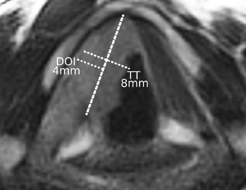
Even more critically, apart from the quantification of vocal cord movement and its impairment, the possible aetiology in terms of the extent of neoplastic invasion into the TAM is debated and has never been standardised in terms of the amount of radiologic and/or histopathologic invasion 22. Given that videoendoscopic assessment of vocal cord mobility is difficult to standardise because of the lack of objective interpretation, radiologic imaging could play a more reproducible role. Traditionally, computed tomography (CT) has been considered the workhorse in laryngeal imaging, reserving magnetic resonance (MR) as a problem solver for specific clinical issues. In fact, the sophistication of scrutiny required for detailed TAM assessment suggests that MR, acquired with optimised high-resolution protocol and surface coils, would be, whenever possible, the imaging technique of choice. Acquisition of axial images parallel to the vocal cords is mandatory to accurately visualise the TAM. On MR, contrast enhanced and diffusion weighted images (DWI) offer a reliable assessment of the deep front of tumour invasion. The combination of signals obtained with different MR sequences may accurately differentiate TAM infiltration from inflammatory oedema. Filauro et al. 22 demonstrated a strong correlation between radiologic depth of invasion (rDOI) and pathological DOI (pDOI), with the latter significantly associated with lymph node metastases, lympho-vascular (LVI), and perineural invasion (PNI). Dagan et al. 23 and Son et al. 24 reported a significant association between tumour thickness (TT) and recurrence-free survival (RFS), proposing different cut-offs of 7 and 4 mm, respectively. In this context, it should be emphasised that DOI and TT represent different concepts, as previously demonstrated in oral cavity carcinoma; in particular, TT may overestimate DOI in exophytic lesions and underestimate it in ulcerated tumours (Cover figure).
MR, with its superior contrast resolution and multiparametric imaging capabilities, allows differentiation between the signal intensity of the vocalis muscle and that of the tumour. In this regard, the most relevant sequences are axial and coronal T2-weighted turbo spin-echo and post-contrast 3D sequences. The use of surface coils is strongly recommended since it allows for improvement in spatial resolution and noise reduction 25. On T2-weighted images, the vocalis muscle typically shows a lower signal intensity compared with carcinoma, enabling reliable differentiation; the combined evaluation of axial and coronal planes is likely crucial for accurate rDOI measurement. Post-contrast sequences are also helpful, as they highlight the marked tumour enhancement compared with the relatively poor one of TAM.
CT has lower soft tissue contrast resolution and may therefore result in less accurate rDOI estimation. However, given its widespread availability, a technical adjustment can be adopted to maximise tumour definition, i.e. reducing the tube voltage from the standard 120 to 100 or even 80 keV, and compensating for the increased image noise by increasing tube current. This adjustment does not correlate to a significant increase in radiation dose. The use of dual-energy CT systems may further improve contrast between tumour and vocalis muscle through spectral imaging, allowing the generation of low-keV monoenergetic images. Finally, emerging photon-counting CT technology may substantially improve diagnostic performance by reducing image noise and providing intrinsic spectral imaging; however, extensive data on this application in head and neck imaging are currently lacking.
Accurate rDOI measurement requires an appropriate para-axial imaging plane aligned with the glottis, obtained by either multiplanar reconstruction of CT datasets or by correct orientation of MR sequences. A reference line should be drawn passing through the anterior commissure and the vocal process of the arytenoid (which is typically aligned with the plane of the disc spaces between C4-C5 and C5-C6); rDOI should then be measured orthogonally from this line to the point of maximal tumour infiltration. In lesions extending cranially toward the ventricle or caudally along the inferior surface of the vocal cord (curved geometry), coronal plane assessment may be preferable. In such cases, the reference line should be drawn tangentially to an ideal reconstruction of the normal profile of the laryngeal mucosa (based on the contralateral side), and rDOI should be calculated orthogonally and deeply from this line (Figs. 1-2).
Given that staging based on vocal cord mobility assessment has been shown to be inherently subjective and operator-dependent, incorporating objective radiological measurements may help overcome this limitation and make clinical staging as uniform as possible across treatment settings, irrespective of histopathological parameters available only in surgically treated patients. In this perspective, imaging-based assessment of TAM involvement and rDOI may provide a more reproducible framework for distinguishing T1-T2a from T2b-T3 diseases, particularly in non-surgical cohorts lacking pathological confirmation.
On the other hand, how to translate these subtle radiologic assessments into a precise and reproducible post-excisional evaluation that is able to establish a histopathologic correlation between cT2a/cT2b and pT2a/pT2b is still matter of ongoing speculation in a multi-institutional effort designed to build a common language between clinicians, radiologists, and surgical pathologists 26.
The thyroarytenoid muscle and its involvement by glottic cancer
In terms of pT classification, one should consider that the entire vocal cord (composed of epithelium, superficial, intermediate and deep layers of lamina propria, TAM, and soft tissues forming the PGS) has an overall thickness ranging from 3.7 (at the level of the anterior paracommissural section) to 7.5 mm (posterior section tangential to the arytenoid vocal process) 27. Little less than half of this thickness consists of the sum of epithelium and vocal ligament, while the lateral part is composed of the TAM and PGS. The literature provides variable reference values concerning TAM thickness: Tayama et al. 28 reported an average of 4 mm, consistent with the above-mentioned values, while the measurements by Hamdan et al.29 and Alonso et al.30 (6.8 and 5.9 mm in males and females, respectively) are at the upper level of the most common estimates. Although the overall thickness of the vocal fold varies along the anteroposterior axis, largely because of differences in the relative contribution of the PGS, the TAM at the glottic level appears to maintain a relatively constant thickness. For this reason, measuring DOI from the superficial to the deepest point of the TAM may provide a reproducible parameter of measure, thereby minimising the potential bias introduced by regional variations in total vocal fold thickness.
Hirano and coworkers 31 empirically defined that vocal cord hypomobility resulting from TAM infiltration arises from involvement of one- to three-fourths of this muscle thickness (and therefore around 1-2 mm). Thus, the vast majority of T1a glottic cancers (limited to one vocal fold with normal cord mobility) that do not extend to the TAM, but only infiltrate the epithelium and vocal ligament, have a DOI ≤ 2 mm. Conversely, morphometric studies have documented that T2 glottic cancers with vocal cord hypomobility have a DOI > 2 mm 32. However, due to technical difficulties in measuring DOI within ulcerated and/or exophytic tumours, it seems more relevant to use a different concept and measure, i.e. the depth of intramuscular invasion (DOIMI) (Fig. 3). Therefore, based on the observations made by Hirano et al. 31, clinical vocal cord hypomobility correlates to a histopathological DOIMI from 1 to 2 mm.
From a biological point of view, muscle invasion in patients with cancer is associated with destructive growth, functional impairment, and poorer prognosis. Skeletal muscle is in fact organised into extracellular matrix-rich surfaces – the epimysium, perimysium, and endomysium – which create cleft-like confined spaces along the interface between dynamic muscle fibres. These structures provide both molecular and physical cues that guide migrating cancer cells, potentially contributing to cancer progression 33. In fact, DOIMI already defines T-subcategories in many other malignancies such as bladder, colorectal, and uterine myometrial cancer, due to its well-known association with decreased overall survival (OS).
In the head and neck region, the extent of DOIMI has only recently gained attention. In fact, DOI was incorporated into the definition of T category for oral cancer only in the 8TNM 12. Since then, multiple studies have confirmed that DOI in tongue cancer is significantly associated with reduced 5-year OS and increased risk of occult metastasis 34,35, contributing to the distinction between T1, T2, and T3 oral tumours. In contrast, within other areas of the head and neck, comparable data remain limited.
In laryngeal cancer, even though the extent of TAM involvement correlates with the severity of mobility impairment 31, this clinical sign can also result from other factors such as a bulky exophytic lesion that impairs vocal cord mobility by its sheer weight. Other mechanisms for impaired vocal cord mobility include supraglottic and hypopharyngeal tumours that secondarily involve the arytenoid cartilage or upper PGS, or infiltrate other intrinsic muscles. Katılmıs et al. 36 analysed whole-organ sections from 133 total laryngectomy (TL) specimens (T2-T4 lesions) and correlated mobility with infiltration patterns. Among 37 patients with vocal cord hypomobility, 23 (62%) showed TAM infiltration, 7 (19%) had posterior crico-arytenoid muscle involvement, and 3 (8%) demonstrated CAU invasion. However, pure vocal cord hypomobility (cT2) should be always clearly distinguished from arytenoid fixation (cT3) as previously highlighted by Succo and coworkers 37 in a series of patients that were treated by open partial horizontal laryngectomies (OPHL), induction chemotherapy plus RT, or TL. These findings again underscore the heterogeneous mechanisms underlying vocal cord hypomobility and suggest the need for a refined diagnostic work-up by endoscopy and imaging to allow for a critical correlation to the specific histopathologic scenario.
Based on the above considerations, radiological and histological correlation of the DOIMI in T2 glottic cancer could serve as a key prognostic indicator (distinguishing T2a from T2b) and may in the future substitute the highly subjective endoscopic finding of vocal cord mobility on which the present 8TNM staging system is still based 12.
The oncologic impact of thyroarytenoid muscle invasion in surgical series
The predominance of RT-based series distinguishing T2 with normal vs altered vocal cord mobility reflects the reliance of non-surgical treatments on clinical and functional surrogates of tumour extent. In the absence of pathological evaluation, accurate identification of a compromised mobility assumes a central prognostic role in cohorts undergoing RT, as it indirectly reflects deeper TAM or PGS infiltration associated with reduced local control. In surgical series, in contrast, the anatomical substrate underlying compromised mobility is addressed directly by compartmental resection, thereby attenuating the prognostic impact of the distinction between cT2a and cT2b. Consequently, pathology-based parameters, such as TAM invasion and DOI (or DOIMI), tend to replace the functional subclassification in surgically-treated populations. This reflects the relative lack of large, homogeneous surgical datasets addressing this distinction.
The prognostic relevance of subdividing T2 glottic carcinoma has been debated for decades, with early observations dating back to 1967 suggesting that vocal fold mobility may represent a clinically meaningful indicator of tumour aggressiveness 9. This historical framework paved the way for progressively refining staging criteria based on functional surrogates of anatomical tumour spread. Building on these observations, in 1997 Chevalier et al. 38 provided an anatomic explanation for the correlation between vocal fold mobility and the depth of tumour infiltration. In their series of 112 patients treated by OPHL type IIa according to the European Laryngological Society classification 39, impaired vocal fold mobility corresponded to partial infiltration of the TAM as defined by Hirano in 1991 31, whereas complete fixation was associated with more than 75% muscle invasion or unequivocal PGS involvement. Although these anatomical differences map closely onto the modern distinction between T2 (impaired mobility) and T3 (fixation), oncologic outcomes were comparable under an OPHL type IIa resection: 5-year local control was 94.4% for impaired mobility and 95.4% for fixation, while cause-specific survival reached 96% and 94.1%, respectively. These findings demonstrate that surgery can mitigate the adverse prognostic implication of deeper invasion but, anatomically, impaired mobility reliably marks TAM infiltration, whereas fixation is consistent with posterior PGS extension, which is the true watershed between T2 and T3 disease.
Over the years, several studies have investigated the prognostic impact of vocal cord mobility, TAM infiltration, and DOI within the spectrum of early-to-intermediate stage glottic tumours. For example, TAM invasion was found to be a negative prognostic factor in surgical cohorts composed of early-stage (T1–T2) glottic cancers studied by Mortuaire et al. 40 and Charbonnier et al. 41. In both series, infiltration of TAM was significantly associated with reduced DFS (p = 0.001 and p = 0.004, respectively), supporting the concept that deeper muscular infiltration may be an adverse biological feature even within early glottic carcinoma.
Parallel to these observations, the concept of DOI began to attract progressive attention. Yilmaz et al. 42 provided one of the first histological demonstrations of the prognostic implications of DOI. In a cohort of 94 surgically-treated patients with T1-T3 tumours, the mean DOI increased significantly with advancing T category, measuring 5.8 mm in T1, 7.2 mm in T2, and 10.1 mm in T3. A DOI of 3.2 mm or greater (and therefore involving the TAM) was consistently associated with a significantly higher rate of cervical metastases, both clinically and pathologically, and DOI independently predicted DFS (p = 0.047).
Despite its recognised importance in other head and neck subsites, DOI was never incorporated into laryngeal staging, likely owing to the complex 3D anatomy of the organ. More recent studies, however, have revisited this limitation. Wang et al. 43 demonstrated that DOI is an independent predictor of both OS and RFS in 412 patients with laryngeal carcinoma and undergoing surgery. DOI thresholds of 10.5 and 19.9 mm stratified tumours into distinct prognostic categories, with an intermediate DOI associated with a 2.6-fold increased risk of mortality and a high DOI with more than a 5-fold increase.
Filauro et al. 22 also evaluated the prognostic role of DOI in 91 patients with surgically-treated glottic tumours. Pathological DOI increased consistently across all T categories, with median values of 1.5-1.2 mm in pT1a-b, 5.3 mm in pT2, 6 mm in pT3, and 14 mm in pT4a. Higher DOI was strongly correlated with adverse pathological features, including PNI, LVI, and nodal metastasis (all p < 0.001). A DOI threshold of 7 mm predicted nodal involvement with an area under the curve (AUC) of 0.82, and DOI significantly affected DFS in univariate analysis.
Ambrosch 44 reported on her experience with TOLMS for 167 moderately advanced glottic carcinomas causing impaired vocal cord mobility (n = 97) or fixation (n = 70). The Kaplan-Meier 5-year local control rate was 74% for pT2 and 68% for pT3. The salvage TL rates were 13.4% and 14.3%, respectively. The 5-year ultimate local control and DFS rates were 87% and 62% in both groups, respectively. This study showed that the prognosis of patients with T2 tumours with compromised motility is quite similar to those harbouring T3 lesions.
Davis et al. 45 evaluated the role of endoscopic vertical partial laryngectomy (EVPL) in a series of 26 patients with T1b-T2 disease. Tumours associated with impaired mobility were managed with EVPL followed by planned postoperative RT, whereas T2 lesions with preserved mobility were treated by surgery alone. Overall local control reached 92.3%, with a 100% local control rate in patients treated with EVPL alone, and 84.5% in those with impaired mobility requiring adjuvant RT. All local recurrences were successfully salvaged by TL, resulting in an OS of 88.5%. Notably, tumours with impaired mobility showed a higher rate of intraoperative upstaging and were more frequently associated with deeper lateral extension toward the TAM and PGS, supporting the concept that reduced vocal cord mobility identifies a biologically distinct subset of T2 tumours requiring wider surgical resection and adjuvant treatment.
In 2014, Canis et al. 46 published a large data set on patients with surgically-treated T2 glottic cancers. Among 391 cases treated by TOLMS, 142 were pT2 with normal vocal cord mobility, 127 pT2 with impaired mobility, and 122 pT3. Five-year OS, RFS, and DSS rates were 72.2%, 76.4%, and 93.2% for the first group, 64.9%, 57.3%, and 83.9% for the second, and 58.6%, 57.8%, and 84.1% for the third. Local control rates at 60 months were 83%, 67.5%, and 71.5%, respectively. Tumours with impaired mobility behaved biologically and prognostically more like T3 than T2 with normal mobility, with no significant differences between T2 with impaired mobility and T3 in several oncologic metrics.
Along the same lines, in 2015, Gorphe et al. 47 analysed a cohort of 148 T2N0 patients, reporting that normal mobility was consistently associated with superior outcomes compared to compromised vocal cord motion. Five-year DFS decreased from 75.5% in the first group to 50.1% in the second, while local control declined from 87.5% to 72.7%. The most dramatic difference was seen in laryngectomy-free survival (LFS), falling from 77.7% to 46.5%. OS also decreased markedly (from 88.6% to 65.6%). Impaired mobility emerged as a significant predictor of poor prognosis, with hazard ratios (HR) ranging from 0.28 to 0.45. Upstaging to T3 occurred in 20% of T2 tumours with impaired mobility, but in only 2% of those without.
In 2017, Day et al. 48 analysed 90 consecutive patients treated by TOLMS, grouping them into Tis, T1, T2 with preserved motility vs T2 with impaired mobility, T3, and T4a. Their results revealed a clear stepwise deterioration in oncologic outcomes once vocal cord mobility was compromised. The first group of patients achieved 5-year local control of 86%, DFS of 65%, DSS of 96%, and laryngeal preservation of 98%, whereas the second experienced significantly inferior outcomes, with local control of 67%, DFS of 45%, DSS of 59%, and laryngeal preservation of 80%. When specifically examining pT2 subcategories, those with normal vocal cord mobility maintained a high local control rate of 88%, whereas those with impaired motion showed a reduction to 78%, accompanied by a greater likelihood of local recurrence and an increased need for TL.
The systematic review conducted by Hendriksma and coworkers 49 qualitatively reinforces the clinical and prognostic importance of distinguishing between T2 with normal vs those with impaired vocal cord mobility, as the latter scenario, related to greater DOI, was consistently associated with worse oncological outcomes in the studies included.
In 2018, Piazza et al. 3 further elucidated the prognostic significance of subdividing pT2 glottic carcinomas based on DOI. Subcategory III, including pT2 extending superficially to the supra- and/or subglottis, demonstrated a 5-year RFS of 81.6% and local control of 90.7%, while subcategory IV, defined by TAM infiltration, yielded RFS of 68.1% and local control of 86.1%. Organ preservation remained high in both groups (95.1% vs 94.4%), but the hazard ratio (HR) for recurrence increased from 3.1 to 4.5 in the presence of TAM infiltration. More recently, the isoprognostic zones defined by Piazza et al. 3 were validated by Marchi et al. 50 in a larger multi-institutional cohort of 637 patients treated by TOLMS. Subcategory III showed a 5-year DSS of 96%, local control of 90%, and laryngeal preservation of 94%, while subcategory IV yielded a 5-year DSS of 92%, 86% local control, and 92% laryngeal preservation. Although primary outcomes were similar, recurrences in subcategory IV were more difficult to treat conservatively: 44% ultimately required TL compared with 31% in subcategory III. These data were subsequently confirmed in another independent data set of 661 patients treated by TOLMS in a retrospective series by Chu et al. 6.
Son et al. 24 examined a cohort of 73 patients treated by TOLMS. In their cohort, including 17 patients with T2 disease, impaired vocal cord mobility was observed in 16.4% of cases, which was one of the most powerful predictors of local recurrence. In particular, impaired vocal cord mobility (HR 8.5; 95% CI 1.45-49.2; p = 0.02) and histopathological tumour thickness (> 4 mm) (HR 6; p = 0.02) were predictive of RFS. Five-year RFS fell from 87.6% in patients with normal mobility to 41.8% in those with reduced mobility, a difference that remained highly significant in multivariate analysis (HR 8.48; 95% CI 1.46-49.16; p = 0.017), thus reinforcing the prognostic significance of vocal cord mobility as a clinical surrogate for deep muscle infiltration.
In 2019, Forner et al. 51 demonstrated significant differences in 5-year local and loco-regional control rates between patients affected by T2 with normal vs impaired vocal cord mobility treated by TOLMS (75.2% vs 57%, p = 0.022). Additionally, 5-year ultimate local control with repeat laser procedures was significantly better for T2 tumours with superficial compared to those with deep extension (87.9% vs 68.1%, p = 0.008). Distant metastases to lungs and liver were encountered in 3 patients affected by T2 with impaired vocal cord mobility, in the absence of regional lymph node involvement at presentation. Postoperative voice outcomes, even though globally satisfactory and in line with those reported by others 52, were markedly worse in T2 with impaired vocal cord mobility: Voice Handicap Index-10 scores improved significantly after surgery for patients with superficial T2 (18.3 to 6.6, p = 0.000) but not for those with T2 infiltrating the TAM (21.4 to 21.3, p = 0.979). Overall, laryngeal preservation was 94.7%, although 11.5% of T2 with impaired vocal cord mobility required salvage TL.
In the series of 209 patients presented by Lionello et al. 53, TAM involvement was associated with a recurrence rate of 37.9% compared to 8.9% in TAM-negative patients (p = 0.005), and DFS decreased from 76.1 to 47.6 months (p = 0.005). Clinically, all cT2 tumours were associated with impaired vocal cord mobility, and more than half of these (56%) showed invasion of the TAM on final histological examination.
The systematic review by Campo et al. 54 analysed 61 studies on treatment of T2N0 glottic tumours. The analysis included 1,452 patients treated by TOLMS, 3,365 managed by RT and 366 by open partial laryngectomies. The authors reported weighted 5-year local control rates of 75.4% for TOLMS, 75.6% for RT, and 94.4% for open partial laryngectomies, with corresponding laryngeal preservation rates of 87%, 82.4%, and 95.8%, respectively. Across the literature, impaired vocal fold mobility has consistently emerged as a negative prognostic factor, demonstrating worse local control and laryngeal preservation outcomes, a pattern observed for both TOLMS and RT.
Another recent work by Wen et al. 55 analysed a contemporary cohort of 216 surgically treated T2 laryngeal carcinomas and confirmed the prognostic relevance of vocal fold mobility within this category. In their series, impaired vocal fold mobility emerged as a significant negative predictor of OS (HR 0.14, 95% CI 0.04-0.44; p = 0.001), independently of the surgical approach chosen (transoral vs open partial).
The available evidence supports a biological continuum in glottic carcinoma, in which vocal fold mobility, presence or not and degree of TAM invasion, and rDOI as well pDOI represent inter-related markers of progressive tumour spread. Within this framework, it is logical to conclude that T2 glottic tumours are far from homogeneous, and lesions with impaired mobility and TAM infiltration (to be upstaged as T2b) demonstrate a distinct oncologic behaviour compared with superficial tumours with normal mobility and no TAM infiltration (to be maintained as T2a). Refining T classification by integrating depth- and compartment-based parameters has the potential to improve prognostic stratification and guide more tailored therapeutic decision-making.
The oncologic impact of thyroarytenoid muscle invasion in RT series
RT has a long tradition of being used to treat laryngeal cancer, even before the advent of TOLMS and OPHL. McCoul published a meta-analysis of 21 studies totaling 2,540 patients treated with RT from 1950 to 2007 for glottic T2 tumours 56. Overall, 5-year local control rates between 47% and 92% were reported with a better outcome for patients without impaired vocal cord mobility. T2 with normal mobility, in fact, had a 5-year local control rate (± standard deviation) of 76.2% (± 10.2), while the comparative figure for T2 with impaired mobility was 64.4% (± 10.2). This difference reached statistical significance with an odds ratio (OR) of 1.83 (95% CI, 1.52-2.20; p = 0.001). The ultimate local control rate after salvage surgery was also significantly better for T2 with normal compared to T2 with impaired mobility (OR, 1.90; 95% CI, 1.23-2.92; p = 0.005), but OS was similar between the 2 subcategories. All the RT series reported in the above-mentioned meta-analysis typically used conformal 3D RT without delineation of target volume based on a thorough description of disease extent and proper imaging, and with parallel opposed fields and/or oblique wedged fields, potentially leading to a suboptimal dose distribution. Interestingly, the authors strongly recommended the subcategorisation of T2 into T2a vs T2b for inclusion in the Cancer Staging Manual, due to its evident impact on prognostication.
Some authors have tried to explore ways to improve the results in “so-called” T2b glottic tumours. Various prognostic factors such as a longer overall treatment time, fraction size, and total dose have been individualised in a series of patients treated by 3D RT from 1956 to 1995 for T2 glottic cancer 57. Moreover, in a series of patients treated from 2006 to 2013, moderately accelerated IMRT (66-70 Gy in 5.5 weeks) was shown to increase 5-year local control rates to 89%; interestingly, no difference was observed between T2a and T2b tumours 58.
Hyperfractionated (HF) 3D RT has also been used in a rather small series of 35 patients with T2 glottic lesions 59. Compared to once-daily RT, HF 3D RT resulted in better local control for both T2a and T2b tumours. This concept has been tested in a randomised trial conducted from 1996 to 2003 (RTOG 9512), which compared 70 Gy in 35 fractions to 79.6 Gy in 66 fractions twice daily, with the objective of improving the treatment of T2b lesions 11. Two hundred and thirty-nine patients with a median follow-up of 7.9 years were randomised. HF RT resulted in a non-significant increase in 5-year local control compared to standard RT (78% vs 70%). Patients with T2a tumours (n = 148) had non-statistically significant better local control at 5 years compared to T2b (n = 91) (T2a 76.8% vs T2b 70%; HR:1.51, 95% CI 0.93-2.44; p = 0.10). The difference between HF and standard RT was also not significant for T2a vs T2b.
Warner et al. 60 performed a MEDLINE systematic review of patients with T2 glottic cancer treated between 1950 and 2016 with either RT (n = 3,191) or TOLMS (n = 1,156). Patients treated by TOLMS were more recently treated. Weighted average 5-year local control rate reached 75.8% after RT and 77.2% after TOLMS, with a worse outcome for T2b after RT or TOLMS compared to T2a.
Recently, a systematic review was conducted on the risk of laryngeal dysfunction after the use of mild-hypofractionated RT for T1-T2 laryngeal cancers 61. The authors concluded that the risk was rather low, typically below 1%. However, whether the use of such regimens will improve the results of RT for T2 glottic lesions remains unknown.
Limitations of the study
This review is inherently constrained by the retrospective and heterogeneous nature of the available literature, encompassing different treatment eras, imaging protocols, and surgical and RT techniques. The majority of cited series rely on surrogate markers such as impaired vocal fold mobility rather than uniformly defined radiologic or histopathologic measures of TAM invasion. Moreover, the proposed threshold values for DOI and DOIMI have not yet been validated prospectively in multicentric studies and may not be directly generalisable across institutions with varying expertise. Finally, the recommendations proposed herein have not been formally tested within the framework of a revised 9TNM staging system or incorporated into prospective clinical trials, and should therefore be interpreted as a rationale for future validation.
Conclusions
Taken together, evidence from both surgical and non-surgical series supports the existence of a prognostic distinction, within the T2 category, between glottic tumours with preserved and those with impaired vocal cord mobility. Therefore, the concept of vocal cord mobility should shift from a purely mechanical characteristic to a biological proxy identifying tumours with different clinical behaviour. Interestingly, this difference tends to diminish in surgical cohorts treated with more extensive procedures, such as OPHL rather than TOLMS, and in RT cohorts receiving treatment intensification through dose escalation or HF. These findings suggest that the adverse prognostic impact of impaired mobility may be counterbalanced only by treatment escalation, indirectly supporting the concept that these tumours represent a biologically and clinically distinct subgroup. Accordingly, these considerations further strengthen our proposal to subdivide T2 glottic cancer into 2 prognostically meaningful subcategories:
- cT2 glottic tumors with superficial extension to the supra- and/or subglottis, with normal vocal cord mobility and without radiologic evidence of TAM invasion should be subclassified as cT2a;
- cT2 with impaired mobility/fixation of the vocal cord but preserved arytenoid movement and radiologic evidence of TAM involvement (or DOIMI) for more than 1 mm should be subclassified as cT2b.
As a consequence of such a proposal, the pT staging of these tumours would change as follows:
- pT2a would be represented by tumours superficially extending from the vocal cord to the supra- and/or subglottis, without histopathologic evidence of TAM infiltration;
- pT2b would be represented by tumours involving the TAM with a DOIMI more than 1 mm.
Conflict of interest statement
The authors declare no conflict of interest.
Funding
This research did not receive any specific grant from funding agencies in the public, commercial, or not-for-profit sectors.
Author contributions
All authors contributed to the interpretation of the data, critically revised the manuscript for important intellectual content, and approved the final version of the manuscript.
Ethical consideration
Not applicable.
History
Received: June 3, 2026
Accepted: July 27, 2026
Figures and tables
Figure 1. Post-contrast magnetic resonance sequence at the level of the glottic plane of a cT1a with normal vocal cord mobility. The dotted line connects the vocal process of the arytenoid cartilage to the anterior commissure. Depth of invasion (DOI) is measured along a segment orthogonal to this line. Note that the exophytic component is excluded from the measurement.
Figure 2. A) Post-contrast magnetic resonance (MR) imaging at the level of the glottic plane of a cT2 with impaired vocal cord mobility. A dotted line is drawn from the vocal process of the right arytenoid cartilage to the anterior commissure. Depth of invasion (DOI) is measured along an orthogonal segment and involves the full thickness of the vocalis muscle; B) Coronal post-contrast MR. The entire thyroarytenoid muscle – including both the lateral thyroarytenoid (ltam) and vocalis muscles (vm) – is involved. On the contralateral side, the normal vm and ventricle (v) are visible; C) Coronal MR T2-weighted image demonstrating altered signal of the vm (more hyperintense compared to the contralateral side). Note that the fat of the paraglottic space (PGS) is preserved and appears enlarged compared to the contralateral side, consistent with oedema.
Figure 3. Histopathologic picture of a glottic tumour with thyroarytenoid muscle invasion and impaired vocal cord mobility (haematoxylin and eosin staining, original magnification 10x). The blue line represents the interface between the vocal ligament and vocalis muscle, the green one the tumour front of invasion, and in red the measure of depth of intramuscular invasion is depicted.
References
- Pittore B, Ismail-Koch H, Davis A. Thyroarytenoid muscle invasion in T1 glottic carcinoma. Eur Arch Otorhinolaryngol. 2009;266:1787-1791. doi:https://doi.org/10.1007/s00405-009-0929-4
- Peretti G, Piazza C, Cocco D. Transoral CO2 laser treatment for T(is)-T(3) glottic cancer: the University of Brescia experience on 595 patients. Head Neck. 2010;32:977-983. doi:https://doi.org/10.1002/hed.21278
- Piazza C, Filauro M, Paderno A. Three-dimensional map of isoprognostic zones in glottic cancer treated by transoral laser microsurgery as a unimodal treatment strategy. Front Oncol. 2018;8. doi:https://doi.org/10.3389/fonc.2018.00175
- Vilaseca I, Nogués-Sabaté A, Avilés-Jurado F. Factors of local recurrence and organ preservation with transoral laser microsurgery in laryngeal carcinomas; CHAID decision-tree analysis. Head Neck. 2019;41:756-764. doi:https://doi.org/10.1002/hed.25422
- Piazza C, Paderno A, Del Bon F. Long-term oncologic outcomes of 1188 Tis-T2 glottic cancers treated by transoral laser microsurgery. Otolaryngol Head Neck Surg. 2021;165:321-328. doi:https://doi.org/10.1177/0194599820983727
- Chu F, Bandi F, Tagliabue M. Transoral laser microsurgery for glottic cancer: landmark 24-year single-institution experience with 661 patients and validation of the isoprognostic zone system. Oral Oncol. 2026;176. doi:https://doi.org/10.1016/j.oraloncology.2026.107917
- Peretti G, Rodrigo J, Bellini E. Contemporary indications and technical limits of transoral laser microsurgery for laryngeal squamous cell carcinoma. Adv Ther. 2026;43:2356-2370. doi:https://doi.org/10.1007/s12325-026-03564-w
- Peretti G, Piazza C, Mensi M. Endoscopic treatment of cT2 glottic carcinoma: prognostic impact of different pT subcategories. Ann Otol Rhinol Laryngol. 2005;114:579-586. doi:https://doi.org/10.1177/000348940511400801
- Martensson B, Fluur E, Jacobsson F. Aspects on treatment of cancer of the larynx. Ann Otol Rhinol Laryngol. 1967;76:313-329. doi:https://doi.org/10.1177/000348946707600202
- Harwood A, Deboer G. Prognostic factors in T2 glottic cancer. Cancer. 1980;45:991-995. doi:https://doi.org/10.1002/1097-0142(19800301)45:5<991::AID-CNCR2820450526>3.0.CO;2-D
- Trotti A, Zhang Q, Bentzen S. Randomized trial of hyperfractionation versus conventional fractionation in T2 squamous cell carcinoma of the vocal cord (RTOG 9512). Int J Radiat Oncol Biol Phys. 2014;89:958-963. doi:https://doi.org/10.1016/j.ijrobp.2014.04.041
- Amin M, Edge S, Greene F. AJCC Cancer Staging Manual. Springer; 2017.
- Rühle A, Subramani R, Su J. Prognostic value of impaired vocal cord mobility in T2N0 glottic cancer treated with IMRT. Laryngoscope. 2026;136:2605-2613. doi:https://doi.org/10.1002/lary.70424
- Ferrari M, Mularoni F, Taboni S. How reliable is assessment of true vocal cord-arytenoid unit mobility in patients affected by laryngeal cancer? A multi-institutional study on 366 patients from the ARYFIX collaborative group. Oral Oncol. 2024;152. doi:https://doi.org/10.1016/j.oraloncology.2024.106744
- Piazza C, Cocco D, Del Bon F. Narrow band imaging and high definition television in the endoscopic evaluation of upper aero-digestive tract cancer. Acta Otorhinolaryngol Ital. 2011;31:70-75.
- Piazza C, Del Bon F, Peretti G. “Biologic endoscopy”: optimization of upper aerodigestive tract cancer evaluation. Curr Opin Otolaryngol Head Neck Surg. 2011;19:67-76. doi:https://doi.org/10.1097/MOO.0b013e328344b3ed
- Piazza C, Del Bon F, Peretti G. Narrow band imaging in endoscopic evaluation of the larynx. Curr Opin Otolaryngol Head Neck Surg. 2012;20:472-476. doi:https://doi.org/10.1097/MOO.0b013e32835908ac
- Piazza C, Mangili S, Del Bon F. Quantitative analysis of videokymography in normal and pathological vocal folds: a preliminary study. Eur Arch Otorhinolaryngol. 2012;269:207-212. doi:https://doi.org/10.1007/s00405-011-1780-y
- Villani F, Paderno A, Fiorentino M. Classifying vocal folds fixation from endoscopic videos with machine learning. Annu Int Conf IEEE Eng Med Biol Soc. 2023;2023:1-4. doi:https://doi.org/10.1109/EMBCC40787.2023.10340017
- Villani F, Fiorentino M, Federici L. A deep-learning approach for vocal fold pose estimation in videoendoscopy. J Imaging Inform Med. 2026;39:842-852. doi:https://doi.org/10.1007/s10278-025-01431-8
- Agrimi E, Pietrogiacomi F, Fiorini L. Artificial intelligence in otolaryngology: redefining automatic laryngeal paralysis assessment for optimal care. SN Comput Sci. 2026;7. doi:https://doi.org/10.1007/s42979-025-04606-w
- Filauro M, Caprioli S, Lovino Camerino P. Depth of invasion assessment in laryngeal glottic carcinoma: a preoperative imaging approach for prognostication. Laryngoscope. 2024;134:3230-3237. doi:https://doi.org/10.1002/lary.31369
- Dagan O, Perlow A, Shoffel-Havakuk H. Effect of radiological tumor thickness on prognosis of early glottic-squamous cell carcinoma treated with radiation. J Voice. Published online 2024. doi:https://doi.org/10.1016/j.jvoice.2024.02.016
- Son H-J, Lee Y, Ku J. Radiological tumor thickness as a risk factor for local recurrence in early glottic cancer treated with laser cordectomy. Eur Arch Otorhinolaryngol. 2018;275:153-160. doi:https://doi.org/10.1007/s00405-017-4793-3
- Maroldi R, Ravanelli M, Farina D. Magnetic resonance for laryngeal cancer. Curr Opin Otolaryngol Head Neck Surg. 2014;22:131-139. doi:https://doi.org/10.1097/MOO.0000000000000036
- Bellini E, Sampieri C, Chu F. Depth of invasion predicts vocal fold motility and muscle invasion in glottic carcinoma. Laryngoscope. Published online 2026. doi:https://doi.org/10.1002/lary.70784
- Sidlof P, Svec J, Horácek J. Geometry of human vocal folds and glottal channel for mathematical and biomechanical modeling of voice production. J Biomech. 2008;41:985-995. doi:https://doi.org/10.1016/j.jbiomech.2007.12.016
- Tayama N, Chan R, Kaga K. Functional definitions of vocal fold geometry for laryngeal biomechanical modeling. Ann Otol Rhinol Laryngol. 2002;111:83-92. doi:https://doi.org/10.1177/000348940211100114
- Hamdan A, Khalifee E, Ziade G. Sexual dimorphism in laryngeal volumetric measurements using magnetic resonance imaging. Ear Nose Throat J. 2020;99:132-136. doi:https://doi.org/10.1177/0145561319840568
- Alonso V, Chagury A, Hachiya A. Diffusion of aniline blue injected into the thyroarytenoid muscle as a proxy for botulinum toxin injection: an experimental study in cadaver larynges. Int Arch Otorhinolaryngol. 2013;17:315-320. doi:https://doi.org/10.7162/S1809-977720130003000012
- Hirano M, Kurita S, Matsuoka H. Vocal fold fixation in laryngeal carcinomas. Acta Otolaryngol. 1991;111:449-454. doi:https://doi.org/10.3109/00016489109137418
- Fang Q, Wang Y, Zhao X. Vocal fold composition and early glottic carcinoma infiltration. World J Surg Oncol. 2012;10. doi:https://doi.org/10.1186/1477-7819-10-178
- Beunk L, Brown K, Nagtegaal I. Cancer invasion into musculature: mechanics, molecules, and implications. Semin Cell Dev Biol. 2019;93:36-45. doi:https://doi.org/10.1016/j.semcdb.2018.07.014
- Faisal M, Bakar M, Sarwar A. Depth of invasion (DOI) as a predictor of cervical nodal metastasis and local recurrence in early stage squamous cell carcinoma of oral tongue (ESSCOT). PloS One. 2018;13. doi:https://doi.org/10.1371/journal.pone.0202632
- Sood R, Singh C, Panda S. Predictors of recurrence, and the impact of adjuvant radiation therapy on survival in early stage (pT1-2, N0) oral cavity squamous cell carcinoma: a matched pair analysis. Oral Oncol. 2025;168. doi:https://doi.org/10.1016/j.oraloncology.2025.107566
- Katilmis H, Ozturkcan S, Ozdemir I. A clinico-pathological study of laryngeal and hypopharyngeal carcinoma: correlation of cord-arytenoid mobility with histopathologic involvement. Otolaryngol Head Neck Surg. 2007;136:291-295. doi:https://doi.org/10.1016/j.otohns.2006.08.022
- Succo G, Cirillo S, Bertotto I. Arytenoid fixation in laryngeal cancer: radiological pictures and clinical correlations with respect to conservative treatments. Cancers (Basel). 2019;11. doi:https://doi.org/10.3390/cancers11030360
- Chevalier D, Laccourreye O, Brasnu D. Cricohyoidoepiglottopexy for glottic carcinoma with fixation or impaired motion of the true vocal cord: 5-year oncologic results with 112 patients. Ann Otol Rhinol Laryngol. 1997;106:364-369. doi:https://doi.org/10.1177/000348949710600502
- Succo G, Peretti G, Piazza C. Open partial horizontal laryngectomies: a proposal for classification by the working committee on nomenclature of the European Laryngological Society. Eur Arch Otorhinolaryngol. 2014;271:2489-2496. doi:https://doi.org/10.1007/s00405-014-3024-4
- Mortuaire G, Francois J, Wiel E. Local recurrence after CO2 laser cordectomy for early glottic carcinoma. Laryngoscope. 2006;116:101-105. doi:https://doi.org/10.1097/01.mlg.0000184524.23282.74
- Charbonnier Q, Thisse A, Sleghem L. Oncologic outcomes of patients with positive margins after laser cordectomy for T1 and T2 glottic squamous cell carcinoma. Head Neck. 2016;38:1804-1809. doi:https://doi.org/10.1002/hed.24518
- Yilmaz T, Hoşal S, Gedikoglu G. Prognostic significance of depth of invasion in cancer of the larynx. Laryngoscope. 1998;108:764-768. doi:https://doi.org/10.1097/00005537-199805000-00025
- Wang X, Cao K, Guo E. Integrating DOI in T classification improves the predictive performance of laryngeal cancer staging. Cancer Biol Ther. 2023;24. doi:https://doi.org/10.1080/15384047.2023.2169040
- Ambrosch P. The role of laser microsurgery in the treatment of laryngeal cancer. Curr Opin Otolaryngol Head Neck Surg. 2007;15:82-88. doi:https://doi.org/10.1097/MOO.0b013e3280147336
- Davis R, Hadley K, Smith M. Endoscopic vertical partial laryngectomy. Laryngoscope. 2004;114:236-240. doi:https://doi.org/10.1097/00005537-200402000-00012
- Canis M, Martin A, Ihler F. Transoral laser microsurgery in treatment of pT2 and pT3 glottic laryngeal squamous cell carcinoma - results of 391 patients. Head Neck. 2014;36:859-866. doi:https://doi.org/10.1002/hed.23389
- Gorphe P, Blanchard P, Breuskin I. Vocal fold mobility as the main prognostic factor of treatment outcomes and survival in stage II squamous cell carcinomas of the glottic larynx. J Laryngol Otol. 2015;129:903-909. doi:https://doi.org/10.1017/S002221511500184X
- Day A, Sinha P, Nussenbaum B. Management of primary T1-T4 glottic squamous cell carcinoma by transoral laser microsurgery. Laryngoscope. 2017;127:597-604. doi:https://doi.org/10.1002/lary.26207
- Hendriksma M, Heijnen B, Sjögren E. Oncologic and functional outcomes of patients treated with transoral CO2 laser microsurgery or radiotherapy for T2 glottic carcinoma: a systematic review of the literature. Curr Opin Otolaryngol Head Neck Surg. 2018;26:84-93. doi:https://doi.org/10.1097/MOO.0000000000000438
- Marchi F, Del Bon F, Chu F. Refining prognostic subcategories in intermediate-advanced glottic cancer: a multicentric study on 637 patients treated by transoral laser microsurgery. Oral Oncol. 2025;164. doi:https://doi.org/10.1016/j.oraloncology.2025.107264
- Forner D, Rigby M, Hart R. Oncological and functional outcomes following transoral laser microsurgery in patients with T2a vs T2b glottic squamous cell carcinoma. J Otolaryngol Head Neck Surg. 2019;48. doi:https://doi.org/10.1186/s40463-019-0346-7
- Peretti G, Piazza C, Del Bon F. Function preservation using transoral laser surgery for T2-T3 glottic cancer: oncologic, vocal, and swallowing outcomes. Eur Arch Otorhinolaryngol. 2013;270:2275-2281. doi:https://doi.org/10.1007/s00405-013-2461-9
- Lionello M, Bertolin A, Nardello E. Could the infiltration of the thyroarytenoid muscle define the pT2 glottic carcinoma?. Head Neck. 2019;41:3639-3646. doi:https://doi.org/10.1002/hed.25893
- Campo F, Zocchi J, Ralli M. Laser microsurgery versus radiotherapy versus open partial laryngectomy for T2 laryngeal carcinoma: a systematic review of oncological outcomes. Ear Nose Throat J. 2021;100:51S-58S. doi:https://doi.org/10.1177/0145561320928198
- Wen J, Luo Q, Wang Z. Oncologic parity in T2 laryngeal cancer: transoral laser surgery and open partial laryngectomy. Eur Arch Otorhinolaryngol. 2025;282:5765-5777. doi:https://doi.org/10.1007/s00405-025-09735-9
- McCoul E, Har-El G. Meta-analysis of impaired vocal cord mobility as a prognostic factor in T2 glottic carcinoma. Arch Otolaryngol Head Neck Surg. 2009;135:479-486. doi:https://doi.org/10.1001/archoto.2009.47
- Le Q, Fu K, Kroll S. Influence of fraction size, total dose, and overall time on local control of T1-T2 glottic carcinoma. Int J Radiat Oncol Biol Phys. 1997;39:115-126. doi:https://doi.org/10.1016/s0360-3016(97)00284-8
- Rock K, Huang S, Tiong A. Partial laryngeal IMRT for T2N0 glottic cancer: impact of image guidance and radiation therapy intensification. Int J Radiat Oncol Biol Phys. 2018;102:941-949. doi:https://doi.org/10.1016/j.ijrobp.2018.03.034
- Fein D, Mendenhall W, Parsons J. T1-T2 squamous cell carcinoma of the glottic larynx treated with radiotherapy: a multivariate analysis of variables potentially influencing local control. Int J Radiat Oncol Biol Phys. 1993;25:605-611. doi:https://doi.org/10.1016/0360-3016(93)90005-g
- Warner L, Lee K, Homer J. Transoral laser microsurgery versus radiotherapy for T2 glottic squamous cell carcinoma: a systematic review of local control outcomes. Clin Otolaryngol. 2017;42:629-636. doi:https://doi.org/10.1111/coa.12790
- Linden S, Philippens M, Sher D. Risk of dysfunctional larynx after radiotherapy for early-stage glottic laryngeal cancer: a systematic review and meta-analysis. Radiother Oncol. 2026;214. doi:https://doi.org/10.1016/j.radonc.2025.111226
Downloads
License

This work is licensed under a Creative Commons Attribution-NonCommercial-NoDerivatives 4.0 International License.
Copyright
Copyright (c) 2026 Società Italiana di Otorinolaringoiatria e chirurgia cervico facciale
How to Cite
- Abstract viewed - 312 times
- PDF downloaded - 101 times